Datasheet is currently unavailable. Try again or CONTACT US
Herpes Simplex Virus Type II Monoclonal Antibody
Mouse Balb/c Monoclonal DBM15.69 IgG1
Mob542-01
100 µL
Liquid
IHC
Herpesvirus
Mouse Balb/c
Shipping info:
$50.00 to US & $70.00 to Canada for most products. Final costs are calculated at checkout.
Product Details
Anti-Herpes Simplex Virus Type II (HSV2) (MOUSE) Monoclonal Antibody - Mob542-01
Herpes, Herpesvirus, Simplex, HSV, HSV2
Mouse Balb/c
Monoclonal
IgG
Target Details
gD
Herpesvirus
Other
Anti-Herpes Simplex Virus Type II (HSV2) antibody was produced in BALB/C mice by repeated immunizations with Parker strain of herpes simplex virus type 2.
The antibody is supplied as a purified immunoglobulin. This antibody reacts with herpes simplex virus type 2. It is specific for the viral glycoprotein D (gD) protein.
Application Details
IHC
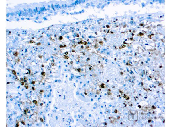
Anti-Herpes Simplex Virus Type II (HSV2) (MOUSE) Monoclonal Antibody was tested with IHC-P positive control infected lung tissues. Cellular Localization: Cytoplasmic, nuclear. The user is advised to validate the use of the products with their tissue specimens prepared and handled in accordance with their laboratory practices. Consult references (Kiernan, 1981: Sheehan & Hrapchak, 1980) for further details on specimen preparation.
Formulation
0.01% (w/v) Sodium Azide
Shipping & Handling
Wet Ice
Store at 2-8°C. This antibody is suitable for use until the expiration date when stored at 2-8°C. Do not use product after the expiration date printed on vial. If reagents are stored under conditions other than those specified here, they must be verified by the user. Diluted reagents should be used promptly. Unused portions of antibody preparation should be discarded after one day. The presence of precipitate or an unusual odor indicates that the antibody is deteriorating and should not be used.
Herpes simplex type 2 (HSV2) belongs to a family that includes HSV1, Epstein-Barr virus (EBV) and Varicella zoster (chicken pox) virus. HSV1 and HSV2 are extremely difficult to distinguish from each other. The HSV-1 strain generally appears in the orafacial organs. HSV2 usually resides in the sacral ganglion at the base of the spine. These viruses have a large DNA genome, capsid, and a lipid bilayer membrane derived from the nuclear membrane of the last host. These viruses are capable of entering a latent phase where the host shows no visible sign of infection. Envelope glycoprotein C (gC) and glycoprotein B (gB) bind to cell surface partials heparin sulfate. Receptor binding protein glycoprotein D (gD) binds to HVEM, nectin-1, and 3-O sulfated heparin sulfate forming complexes with glycoprotein H (gH) and glycoprotein (gL) creating an entry pore for the viral capsid.
This product is for research use only and is not intended for therapeutic or diagnostic applications. Please contact a technical service representative for more information. All products of animal origin manufactured by Rockland Immunochemicals are derived from starting materials of North American origin. Collection was performed in United States Department of Agriculture (USDA) inspected facilities and all materials have been inspected and certified to be free of disease and suitable for exportation. All properties listed are typical characteristics and are not specifications. All suggestions and data are offered in good faith but without guarantee as conditions and methods of use of our products are beyond our control. All claims must be made within 30 days following the date of delivery. The prospective user must determine the suitability of our materials before adopting them on a commercial scale. Suggested uses of our products are not recommendations to use our products in violation of any patent or as a license under any patent of Rockland Immunochemicals, Inc. If you require a commercial license to use this material and do not have one, then return this material, unopened to: Rockland Inc., P.O. BOX 5199, Limerick, Pennsylvania, USA.

