Rabbit IgG (H&L) Antibody DyLight™ 800 Conjugated Pre-Adsorbed
Goat Polyclonal
22 References
611-145-122
100 µg
Lyophilized
WB, Dot Blot, Other
Rabbit
Goat
Shipping info:
$50.00 to US & $70.00 to Canada for most products. Final costs are calculated at checkout.
Product Details
Goat Anti-Rabbit IgG (H&L) Antibody DyLight™ 800 Conjugated (Min X Bv Ch Gt GP Ham Hs Hu Ms Rt & Sh Serum Proteins) - 611-145-122
Goat anti-Rabbit IgG Antibody DyLight™800 Conjugation, Goat anti-Rabbit IgG DyLight™ 800 Conjugated Antibody
Goat
IgG (H&L)
DyLight™ 800
Polyclonal
IgG
Target Details
Rabbit
Rabbit IgG whole molecule
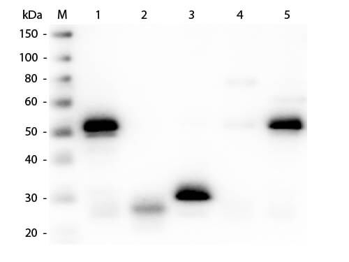
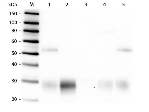
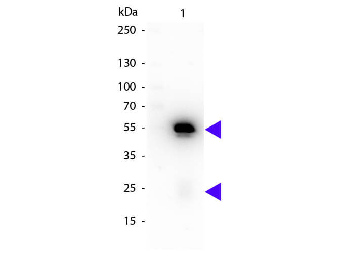
This DyLight™ 800 conjugated secondary antibody was prepared from monospecific antiserum by immunoaffinity chromatography using Rabbit IgG coupled to agarose beads followed by solid phase adsorption(s) to remove any unwanted reactivities. Assay by immunoelectrophoresis resulted in a single precipitin arc against anti-Goat Serum, Rabbit IgG and Rabbit Serum. No reaction was observed against Bovine, Chicken, Goat, Guinea Pig, Hamster, Horse, Human, Mouse, Rat and Sheep Serum Proteins. This antibody will react with heavy chains of rabbit IgG and with light chains of most rabbit immunoglobulins.
Application Details
Dot Blot
Other, WB
- View References
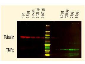
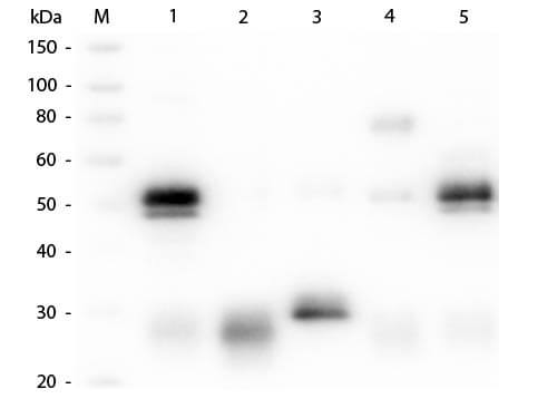
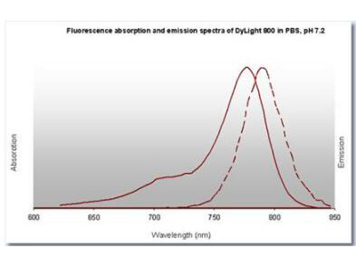
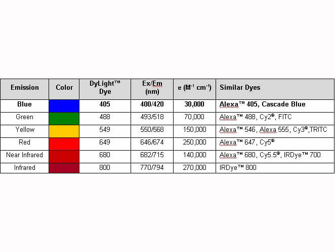
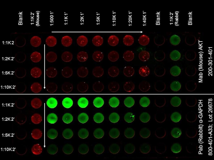
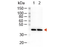
Anti-Rabbit IgG DyLight™800 has been tested by dot blot. The emission spectra for this DyLight™ conjugate match the principle output wavelengths of most common fluorescence instrumentation.
Formulation
1.0 mg/mL by UV absorbance at 280 nm
0.02 M Potassium Phosphate, 0.15 M Sodium Chloride, pH 7.2
0.01% (w/v) Sodium Azide
10 mg/mL Bovine Serum Albumin (BSA) - Immunoglobulin and Protease free
100 µL
Restore with deionized water (or equivalent)
Shipping & Handling
Ambient
Store vial at 4° C prior to restoration. For extended storage aliquot contents and freeze at -20° C or below. Avoid cycles of freezing and thawing. Centrifuge product if not completely clear after standing at room temperature. This product is stable for several weeks at 4° C as an undiluted liquid. Dilute only prior to immediate use.
Expiration date is one (1) year from date of receipt.
Conjugated DyLight™ 800 secondary antibody is designed for immunofluorescence microscopy, fluorescence based plate assays (FLISA) and fluorescent western blotting. This product is also suitable for multiplex analysis, including multicolor imaging, utilizing various commercial platforms.
Shi G et al. (2023). Astragaloside IV promotes cerebral angiogenesis and neurological recovery after focal ischemic stroke in mice via activating PI3K/Akt/mTOR signaling pathway. Heliyon.
Applications
WB, IB, PCA
Szewczyk B et al. (2023). FUS ALS neurons activate major stress pathways and reduce translation as an early protective mechanism against neurodegeneration. Cell Rep.
Applications
WB, IB, PCA
Dabrowski R et al. (2023). Parallel phospholipid transfer by Vps13 and Atg2 determines autophagosome biogenesis dynamics. J Cell Biol.
Applications
WB, IB, PCA
Joshi, H et al. (2022). L-plastin enhances NLRP3 inflammasome assembly and bleomycin-induced lung fibrosis. Cell Reports
Applications
WB, IB, PCA
Wang B et al. (2022). Intracerebral hemorrhage alters α2δ1 and thrombospondin expression in rats. Exp Ther Med.
Applications
WB, IB, PCA
Xue J et al. (2021). Caffeine improves bladder function in diabetic rats via a neuroprotective effect. Exp Ther Med.
Applications
WB, IB, PCA
Zhang P et al. (2021). Atorvastatin alleviates microglia-mediated neuroinflammation via modulating the microbial composition and the intestinal barrier function in ischemic stroke mice. Free Radic Biol Med.
Applications
WB, IB, PCA
Chen J et al. (2021). Inhibition of Acyl-CoA Synthetase Long-Chain Family Member 4 Facilitates Neurological Recovery After Stroke by Regulation Ferroptosis. Front Cell Neurosci.
Applications
WB, IB, PCA
Tichy ED et al. (2021). Persistent NF-κB activation in muscle stem cells induces proliferation-independent telomere shortening. Cell Rep.
Applications
WB, IB, PCA
Navarro R et al. (2020). TGF‐β‐induced IGFBP‐3 is a key paracrine factor from activated pericytes that promotes colorectal cancer cell migration and invasion. Mol Oncol.
Applications
WB, IB, PCA
Mitra S, Bodor DL, David AF, et al. (2020). Genetic screening identifies a SUMO protease dynamically maintaining centromeric chromatin. Nat Commun.
Applications
WB, IB, PCA
Takahashi H, Ranjan A, Chen S, et al. (2020). The role of Mediator and Little Elongation Complex in transcription termination. Nat Commun.
Applications
WB, IB, PCA
Acin-Perez R et al. (2020). Analyzing electron transport chain supercomplexes. Methods Cell Biol.
Applications
WB, IB, PCA
Mastro TL et al. (2020). A sex difference in the response of the rodent postsynaptic density to synGAP haploinsufficiency. Elife.
Applications
WB, IB, PCA
McManus MJ et al. (2019). Mitochondrial DNA variation dictates expressivity and progression of nuclear DNA mutations causing cardiomyopathy. Cell Metab.
Applications
WB, IB, PCA
Latorre-Pellicer A et al. (2019). Regulation of mother-to-offspring transmission of mtDNA heteroplasmy. Cell Metab.
Applications
WB, IB, PCA
Chen J et al. (2019). Ginsenoside Rg1 promotes cerebral angiogenesis via the PI3K/Akt/mTOR signaling pathway in ischemic mice. Eur J Pharmacol.
Applications
WB, IB, PCA
Pereira et al. (2018). mitoTev-TALE: a monomeric DNA editing enzyme to reduce mutant mitochondrial DNA levels. EMBO Molecular Medicine
Applications
WB, IB, PCA
Nguyen et al. (2017). FLT3 activating mutations display differential sensitivity to multiple tyrosine kinase inhibitors. Oncotarget
Applications
WB, IB, PCA
Meyer et al. (2016). Adipocytes promote pancreatic cancer cell proliferation via glutamine transfer. Biochemistry and Biophysics Reports
Applications
WB, IB, PCA
Wu JW et al. (2016). Neuronal activity enhances tau propagation and tau pathology in vivo. Nat Neurosci.
Applications
WB, IB, PCA
Klammer M et al. (2012). Phosphosignature predicts dasatinib response in non-small cell lung cancer. Mol Cell Proteomics.
Applications
WB, IB, PCA
This product is for research use only and is not intended for therapeutic or diagnostic applications. Please contact a technical service representative for more information. All products of animal origin manufactured by Rockland Immunochemicals are derived from starting materials of North American origin. Collection was performed in United States Department of Agriculture (USDA) inspected facilities and all materials have been inspected and certified to be free of disease and suitable for exportation. All properties listed are typical characteristics and are not specifications. All suggestions and data are offered in good faith but without guarantee as conditions and methods of use of our products are beyond our control. All claims must be made within 30 days following the date of delivery. The prospective user must determine the suitability of our materials before adopting them on a commercial scale. Suggested uses of our products are not recommendations to use our products in violation of any patent or as a license under any patent of Rockland Immunochemicals, Inc. If you require a commercial license to use this material and do not have one, then return this material, unopened to: Rockland Inc., P.O. BOX 5199, Limerick, Pennsylvania, USA.

