Datasheet is currently unavailable. Try again or CONTACT US
RFP Antibody Fluorescein Conjugated Pre-Adsorbed
Rabbit Polyclonal
4 References
600-402-379
100 µg
Lyophilized
IHC, IF, Dot Blot, FISH
RFP, rRFP
Rabbit
Shipping info:
$50.00 to US & $70.00 to Canada for most products. Final costs are calculated at checkout.
Product Details
Anti-RFP (RABBIT) Antibody Fluorescein Conjugated (Min X Hu Ms & Rt Serum Proteins) - 600-402-379
rabbit anti-RFP antibody fluorescein conjugation, FITC conjugated rabbit anti-RFP antibody, DsRed, rDsRed, Discosoma sp. Red Fluorescent Protein, Red fluorescent protein drFP583
Rabbit
Fluorescein (FITC)
Polyclonal
IgG
Target Details
DsRed - View All DsRed Products
RFP, rRFP
Recombinant Protein
The immunogen is a Red Fluorescent Protein (RFP) fusion protein corresponding to the full length amino acid sequence (234aa) derived from the mushroom polyp coral Discosoma.
This product was prepared from monospecific antiserum by immunoaffinity chromatography using Red Fluorescent Protein (Discosoma) coupled to agarose beads followed by solid phase adsorption(s) to remove any unwanted reactivities. Expect reactivity against RFP and its variants: mCherry, tdTomato, mBanana, mOrange, mPlum, mOrange and mStrawberry. Assay by immunoelectrophoresis resulted in a single precipitin arc against anti-fluorescein, anti-Rabbit Serum and purified and partially purified Red Fluorescent Protein (Discosoma). No reaction was observed against Human, Mouse or Rat serum proteins. ELISA was used to confirm specificity at less than 0.1% of target signal.
Q9U6Y8 - UniProtKB
Application Details
Dot Blot
FISH, IF, IHC
- View References
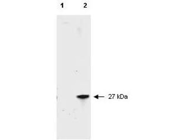
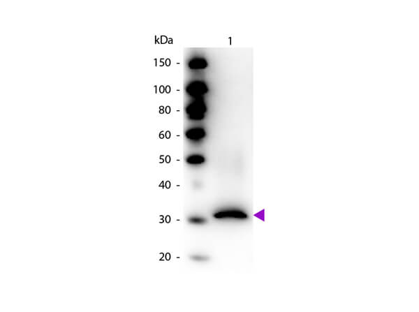
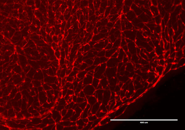
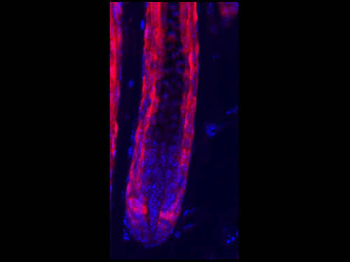
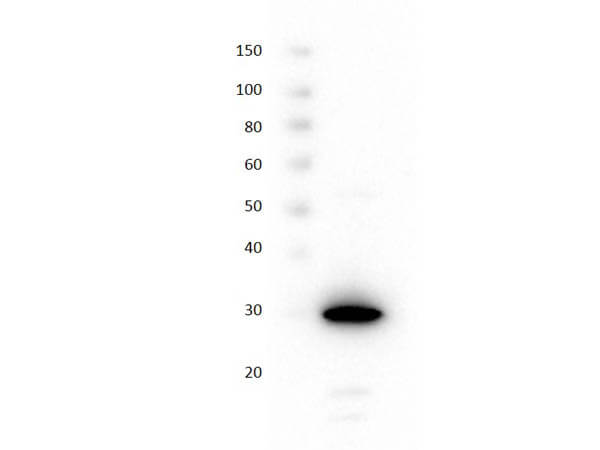
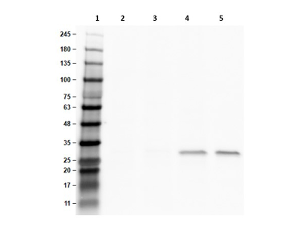
Polyclonal anti-RFP is designed to detect RFP and its variants. This fluorescein conjugated antibody has been tested by dot blot and can be used to detect RFP by ELISA (sandwich or capture) for the direct binding of antigen. Significant amplification of signal is achieved using fluorochrome conjugated polyclonal anti-RFP relative to the fluorescence of RFP alone. Optimal titers for applications should be determined by the researcher.
Formulation
1.0 mg/mL by UV absorbance at 280 nm
0.02 M Potassium Phosphate, 0.15 M Sodium Chloride, pH 7.2
0.01% (w/v) Sodium Azide
10 mg/mL Bovine Serum Albumin (BSA) - Immunoglobulin and Protease free
100 µL
Restore with deionized water (or equivalent)
Shipping & Handling
Ambient
Store vial at 4° C prior to restoration. For extended storage aliquot contents and freeze at -20° C or below. Avoid cycles of freezing and thawing. Centrifuge product if not completely clear after standing at room temperature. This product is stable for several weeks at 4° C as an undiluted liquid. Dilute only prior to immediate use.
Expiration date is one (1) year from date of receipt.
Fluorescent proteins such as Discosoma Red Fluorescent Protein (DsRed) from sea anemone Discosoma sp. mushroom or green fluorescent protein (GFP) from Aequorea victoria jellyfish are widely used in research practice. Fusion RFP and GFP commonly serve as marker for gene expression and protein localization. As DsRed and GFP share only 19% identity, therefore, in general, anti-GFP antibodies do not recognize DsRed protein and vice versa. Structurally, Discosoma red fluorescent protein is similar to Aequorea green fluorescent protein in terms of its overall fold (a β-can) and chromophore-formation chemistry. However, Discosoma red fluorescent protein undergoes an additional step in the chromophore maturation and obligates tetrameric structure. Rockland offers many controls, monoclonal, and polyclonal antibodies for RFP.
Wang SB et al. (2021). Retinal and callosal activity-dependent chandelier cell elimination shapes binocularity in primary visual cortex. Neuron.
Applications
IHC, ICC, Histology
Childs BG et al. (2021). Senescent cells suppress innate smooth muscle cell repair functions in atherosclerosis. Nat Aging.
Applications
IF, Confocal Microscopy
Mevel R et al. (2020). RUNX1 marks a luminal castration-resistant lineage established at the onset of prostate development. Elife.
Applications
IF, Confocal Microscopy; IHC, ICC, Histology
Yokota T, McCourt J, Ma F, et al. (2020). Type V Collagen in Scar Tissue Regulates the Size of Scar after Heart Injury. Cell.
Applications
ISH, FISH
This product is for research use only and is not intended for therapeutic or diagnostic applications. Please contact a technical service representative for more information. All products of animal origin manufactured by Rockland Immunochemicals are derived from starting materials of North American origin. Collection was performed in United States Department of Agriculture (USDA) inspected facilities and all materials have been inspected and certified to be free of disease and suitable for exportation. All properties listed are typical characteristics and are not specifications. All suggestions and data are offered in good faith but without guarantee as conditions and methods of use of our products are beyond our control. All claims must be made within 30 days following the date of delivery. The prospective user must determine the suitability of our materials before adopting them on a commercial scale. Suggested uses of our products are not recommendations to use our products in violation of any patent or as a license under any patent of Rockland Immunochemicals, Inc. If you require a commercial license to use this material and do not have one, then return this material, unopened to: Rockland Inc., P.O. BOX 5199, Limerick, Pennsylvania, USA.

