Datasheet is currently unavailable. Try again or CONTACT US
Myosin phospho S19/phospho S20 Antibody
Rabbit Polyclonal
12 References
600-401-416S
600-401-416
25 µL
100 µg
Liquid (sterile filtered)
Liquid (sterile filtered)
WB, ELISA, IHC, IF, Multiplex
Human, Mouse
Rabbit
Shipping info:
$50.00 to US & $70.00 to Canada for most products. Final costs are calculated at checkout.
Product Details
Anti-Myosin pS19/pS20 (RABBIT) Antibody - 600-401-416
rabbit anti-Myosin p19/pS20 antibody, Myosin regulatory light chain 12A, Myosin regulatory light chain MRLC3, Myosin regulatory light chain 2 nonsarcomeric, Myosin RLC, MLC-2B, HEL-S-24, Epididymis secretory protein Li 24, MLCB
Rabbit
Polyclonal
IgG
Target Details
MYL12A - View All MYL12A Products
Human, Mouse
Phosphorylation
Conjugated Peptide
Human Myosin Light Chain phospho peptide corresponding to a region near the amino terminus of the human smooth/non-muscle form of myosin regulatory light chain conjugated to Keyhole Limpet Hemocyanin (KLH).
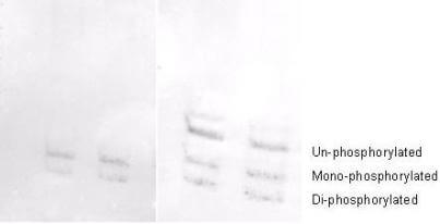
This affinity purified antibody is directed against the regulatory light chain of smooth and non-muscle myosin. The antibody is phosphospecific and detects monophosphorylated and diphosphorylated forms of the protein. The product was affinity purified from monospecific antiserum by immunoaffinity purification. Antiserum was first purified against the phosphorylated form of the immunizing peptide. The resultant affinity purified antibody was then cross-adsorbed against the non-phosphorylated form of the immunizing peptide. This phosphospecific polyclonal antibody is specific for the phosphorylated pS19/pS20 form of the protein, depending on the source origin of the protein. Reactivity with non-phosphorylated myosin light chain is less than 1% by ELISA. Cross reactivity is expected with myosin light chain from human and mouse. Reactivity with the protein from other species has not been determined. However, the sequence of the immunogen is nearly identical in mammalian and avian species. BLAST search analysis was used to determine that the smooth and non-muscle forms of myosin regulatory light chain have identical sequences. Cross reactivity is expected.
Application Details
ELISA, IHC, WB
IF, Multiplex
- View References
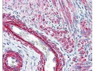
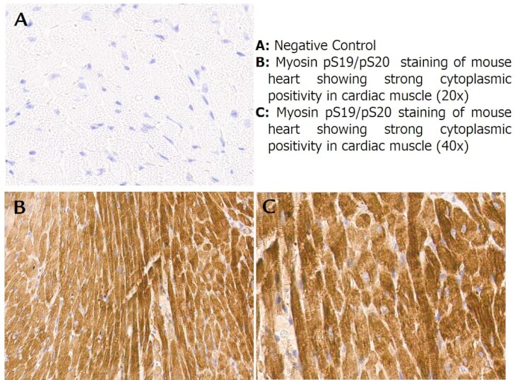
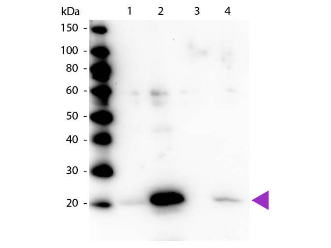
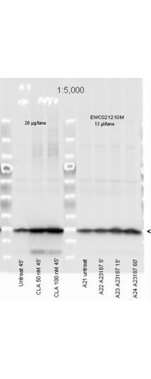
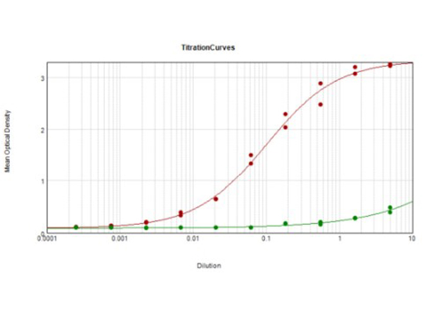
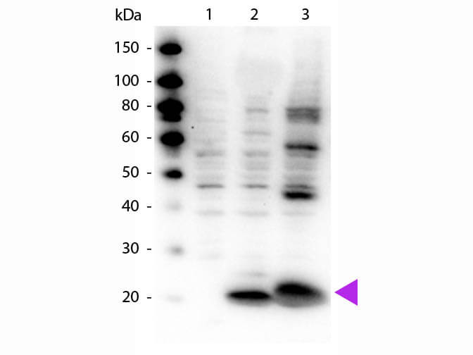
Rabbit Anti-Myosin pS19/pS20 Antibody was tested by ELISA, immunohistochemistry, and western blotting. Immunoblotting was used to show reactivity with unstimulated and stimulated cardiac myocytes, 3T3 whole cell lysates, and regulatory light chain and smooth muscle phospho recombinant protein. The antibody was also reactive with the phosphorylated form of the immunizing peptide and minimally reactive with the non-phosphorylated form of the immunizing peptide. Although not tested, this antibody is likely functional by immunoprecipitation.
Formulation
1.19 mg/mL by UV absorbance at 280 nm
0.02 M Potassium Phosphate, 0.15 M Sodium Chloride, pH 7.2
0.01% (w/v) Sodium Azide
None
Shipping & Handling
Dry Ice
Store vial at -20° C prior to opening. Aliquot contents and freeze at -20° C or below for extended storage. Avoid cycles of freezing and thawing. Centrifuge product if not completely clear after standing at room temperature. This product is stable for several weeks at 4° C as an undiluted liquid. Dilute only prior to immediate use.
Expiration date is one (1) year from date of receipt.
Myosin is the major component of thick muscle filaments, and is a long asymmetric molecule containing a globular head and a long tail. The molecule consists of two heavy chains each ~200,000 daltons, and four light chains each ~16,000 - 21,000 daltons. Activation of smooth and cardiac muscle primarily involves pathways that increase calcium levels and myosin phosphorylation, resulting in contraction. Myosin light chain phosphatase acts to regulate muscle contraction by dephosphorylating activated myosin light chain. This antibody is specific for the phosphorylated form of myosin light chain. The selected peptide sequence used to generate the polyclonal antibody is located near the amino terminal end of the polypeptide corresponding to the smooth/non-muscle form of myosin regulatory light chain found in cardiac myocytes in addition to smooth and non-muscle cells. This sequence differs from that of the sarcomeric/cardiac form of myosin regulatory light chain that has a different sequence around the phosphorylation site. Human and mouse have almost identical sequences. In human the phosphorylation site is pS19, while in mouse the site maps to pS20. Myosin may play a role in disorders such as cardiomyopathies. Anti-Myosin pS19/sP20 Antibody is useful for researcher interested in stem cell and enzyme researcher.
Putz S et al. (2021). Caldesmon ablation in mice causes umbilical herniation and alters contractility of fetal urinary bladder smooth muscle. J Gen Physiol
Applications
WB, IB, PCA
Harbom LJ et al. (2019). The effect of rho kinase inhibition on morphological and electrophysiological maturity in iPSC-derived neurons. Cell Tissue Res.
Applications
WB, IB, PCA
Salomon et al. (2017). Contractile forces at tricellular contacts modulate epithelial organization and monolayer integrity. Nature Communications
Applications
IF, Confocal Microscopy; WB, IB, PCA
Panousopoulou et al. (2016). Epiboly generates the epidermal basal monolayer and spreads the nascent mammalian skin to enclose the embryonic body. Journal of Cell Science
Applications
IHC, ICC, Histology
Sánchez-Elordi, E., Vicente-Manzanares, M., Díaz, E., Legaz, M. E., & Vicente, C. (2016). Plant–pathogen interactions: Sugarcane glycoproteins induce chemotaxis of smut teliospores by cyclic contraction and relaxation of the cytoskeleton. South African Journal of Botany
Applications
WB, IB, PCA
Solanes et al. (2015). Space exploration by dendritic cells requires maintenance of myosin II activity by IP3 receptor 1. The EMBO Journal
Applications
WB, IB, PCA
Logue et al. (2015). Erk regulation of actin capping and bundling by Eps8 promotes cortex tension and leader bleb-based migration. Elife
Applications
IF, Confocal Microscopy; WB, IB, PCA; Multiplex Assay
Newell-Litwa et al. (2015). ROCK1 and 2 differentially regulate actomyosin organization to drive cell and synaptic polarity. Journal of Cell Biology
Applications
IF, Confocal Microscopy; WB, IB, PCA
Liu YJ et al. (2015). Confinement and low adhesion induce fast amoeboid migration of slow mesenchymal cells. Cell.
Applications
WB, IB, PCA
Roland AB et al. (2014). Cannabinoid-induced actomyosin contractility shapes neuronal morphology and growth. Elife
Applications
IF, Confocal Microscopy
Fabbiano S et al. (2014). Genetic dissection of the vav2-rac1 signaling axis in vascular smooth muscle cells. Mol Cell Biol.
Applications
WB, IB, PCA
Edelson JR, Brautigan DL. (2011). The Discodermia calyx toxin calyculin a enhances cyclin D1 phosphorylation and degradation, and arrests cell cycle progression in human breast cancer cells Toxins (Basel)
Applications
WB, IB, PCA
This product is for research use only and is not intended for therapeutic or diagnostic applications. Please contact a technical service representative for more information. All products of animal origin manufactured by Rockland Immunochemicals are derived from starting materials of North American origin. Collection was performed in United States Department of Agriculture (USDA) inspected facilities and all materials have been inspected and certified to be free of disease and suitable for exportation. All properties listed are typical characteristics and are not specifications. All suggestions and data are offered in good faith but without guarantee as conditions and methods of use of our products are beyond our control. All claims must be made within 30 days following the date of delivery. The prospective user must determine the suitability of our materials before adopting them on a commercial scale. Suggested uses of our products are not recommendations to use our products in violation of any patent or as a license under any patent of Rockland Immunochemicals, Inc. If you require a commercial license to use this material and do not have one, then return this material, unopened to: Rockland Inc., P.O. BOX 5199, Limerick, Pennsylvania, USA.

