Datasheet is currently unavailable. Try again or CONTACT US
Glutamate Dehydrogenase Antibody
Rabbit Polyclonal
6 References
100-4158
2 mL
Lyophilized
WB, Other
Bovine
Rabbit
Shipping info:
$50.00 to US & $70.00 to Canada for most products. Final costs are calculated at checkout.
Product Details
Anti-Glutamate Dehydrogenase (Bovine Liver) (RABBIT) Antibody - 100-4158
rabbit anti-Glutamate Dehydrogenase Antibody, Glutamate dehydrogenase 1 mitochondrial, GDH 1
Rabbit
Polyclonal
Antiserum
Target Details
GLUD1 - View All GLUD1 Products
Bovine
Native Protein
This antibody was prepared from whole rabbit serum produced by repeated immunizations with a full length Glutamate Dehydrogenase protein isolated from Bovine Liver.
This product was prepared from monospecific antiserum by a delipidation and defibrination. Assay by immunoelectrophoresis resulted in a single precipitin arc against anti-rabbit serum, purified and partially purified Glutamate Dehydrogenase [Bovine Liver]. BLAST analysis was used to determine that cross reactivity is suggested for both mitochondrial and brain isoforms (GDH1 and GDH2), from both bovine and human sources. Additionally similar reactivity is suggested for most primate species including green monkey, white gibbon, chimpanzee orangutan, and gorilla. A high degree of sequence homology is also noted for GDH from chicken, mouse, rat, dog, and other mammals as well as Xenopus tropicalis, zebrafish, rainbow trout and Atlantic salmon. Cross reactivity against Glutamate Dehydrogenase from other tissues and species may occur but have not been specifically determined.
Application Details
WB
Other
- View References
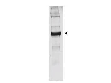
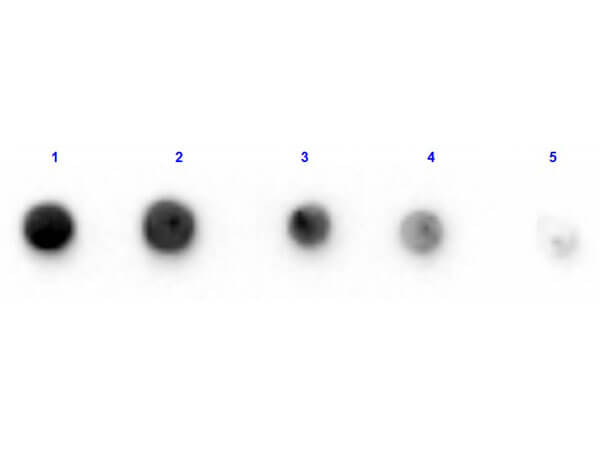
This antibody has been tested by western blot. Specific conditions for reactivity should be optimized by the end user. Bovine glutamate dehydrogenase exists as a homohexamer located within the mitochondrial matrix. Expect a band approximately 56 kDa in size corresponding to glutamate dehydrogenase monomer subunit by western blotting in the appropriate cell or tissue extract. Anti-Glutamate Dehydrogenase Antibody is suitable for use in ELISA.
Formulation
85 mg/mL by Refractometry
0.02 M Potassium Phosphate, 0.15 M Sodium Chloride, pH 7.2
0.01% (w/v) Sodium Azide
None
2.0 mL
Restore with deionized water (or equivalent)
Shipping & Handling
Ambient
Store vial at 4° C prior to restoration. For extended storage aliquot contents and freeze at -20° C or below. Avoid cycles of freezing and thawing. Centrifuge product if not completely clear after standing at room temperature. This product is stable for several weeks at 4° C as an undiluted liquid. Dilute only prior to immediate use.
Expiration date is one (1) year from date of receipt.
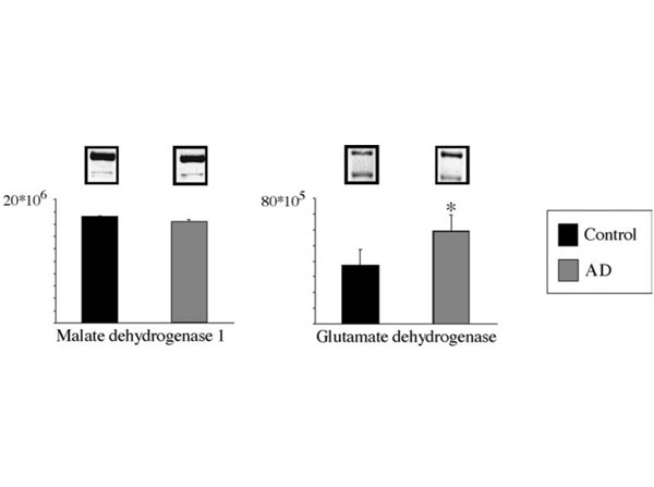
Glutamate is a major excitatory neurotransmitter. One enzyme central to the metabolism of glutamate is glutamate dehydrogenase (GDH1; EC 1.4.1.3), that catalyzes the reversible deamination of L-glutamate to 2-oxoglutarate using NAD+ or NADP+. Mammalian GDH is composed of six identical subunits, and the regulation of GDH is very complex. It has been a major goal to identify the substrate and regulatory binding sites of GDH. It is only in recent years that the three-dimensional structure of GDH from microorganisms is available. Very recently, crystallization of bovine liver GDH was reported for the first time from the mammalian sources. However, remarkably little is known about the detailed structure of mammalian GDH, especially the brain enzymes.
Alam P et al. (2020). Acetazolamide causes renal HCO−3 wasting but inhibits ammoniagenesis and prevents the correction of metabolic acidosis by the kidney. Am J Physiol Renol Physiol.
Applications
WB, IB, PCA
Karaca M et al. (2018). Liver glutamate dehydrogenase controls whole-body energy partitioning through amino acid–derived gluconeogenesis and ammonia homeostasis. Diabetes.
Applications
WB, IB, PCA
Morvay PL et al. (2017). Differential activities of peroxisomes along the mouse intestinal epithelium. Cell Biochem Funct.
Applications
WB, IB, PCA
Zufferey A et al. (2014). Characterization of the platelet granule proteome: evidence of the presence of MHC1 in alpha-granules. J Proteomics.
Applications
WB, IB, PCA
Pougovkina O et al. (2014). Mitochondrial protein acetylation is driven by acetyl-CoA from fatty acid oxidation. Hum Mol Genet.
Applications
WB, IB, PCA
Anthonio EA et al. (2009). Small G proteins in peroxisome biogenesis: the potential involvement of ADP-ribosylation factor 6. AMC Cell Biol.
Applications
WB, IB, PCA
This product is for research use only and is not intended for therapeutic or diagnostic applications. Please contact a technical service representative for more information. All products of animal origin manufactured by Rockland Immunochemicals are derived from starting materials of North American origin. Collection was performed in United States Department of Agriculture (USDA) inspected facilities and all materials have been inspected and certified to be free of disease and suitable for exportation. All properties listed are typical characteristics and are not specifications. All suggestions and data are offered in good faith but without guarantee as conditions and methods of use of our products are beyond our control. All claims must be made within 30 days following the date of delivery. The prospective user must determine the suitability of our materials before adopting them on a commercial scale. Suggested uses of our products are not recommendations to use our products in violation of any patent or as a license under any patent of Rockland Immunochemicals, Inc. If you require a commercial license to use this material and do not have one, then return this material, unopened to: Rockland Inc., P.O. BOX 5199, Limerick, Pennsylvania, USA.


