Datasheet is currently unavailable. Try again or CONTACT US
GFP (GOAT) Antibody Biotin Conjugated
Goat Polyclonal
41 References
600-106-215
1 mg
Lyophilized
WB, ELISA, IHC, IF, FC, EM, IP, Multiplex, Other, Purification
GFP, eGFP, rGFP
Goat
Shipping info:
$50.00 to US & $70.00 to Canada for most products. Final costs are calculated at checkout.
Product Details
Anti-GFP (GOAT) Antibody Biotin Conjugated - 600-106-215
goat anti-GFP Antibody biotin Conjugation, biotin conjugated goat anti-GFP antibody, Green Fluorescent Protein, GFP antibody, Green Fluorescent Protein antibody, EGFP, enhanced Green Fluorescent Protein, Aequorea victoria, Jellyfish
Goat
Biotin
Polyclonal
IgG
Target Details
GFP, eGFP, rGFP
Recombinant Protein
Recombinant Green Fluorescent Protein (GFP) fusion protein corresponding to the full length amino acid sequence (246aa) derived from the jellyfish Aequorea victoria.
Anti-GFP was prepared from monospecific antiserum by immunoaffinity chromatography using Green Fluorescent Protein (Aequorea victoria) coupled to agarose beads followed by solid phase adsorption(s) to remove any unwanted reactivities. Assay by immunoelectrophoresis resulted in a single precipitin arc against anti-Goat Serum, anti-biotin and purified and partially purified Green Fluorescent Protein (Aequorea victoria) Serum. No reaction was observed against Human, Mouse and Rat Serum Proteins.
P42212 - UniProtKB
Application Details
ELISA, WB
EM, FC, IF, IHC, IP, Other, Purification, Multiplex
- View References
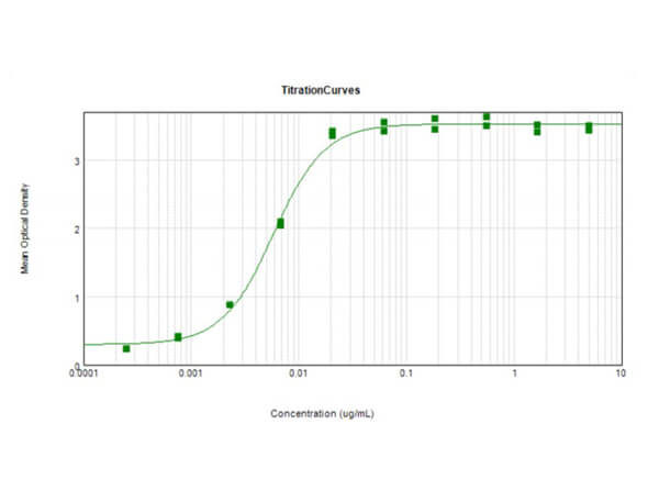
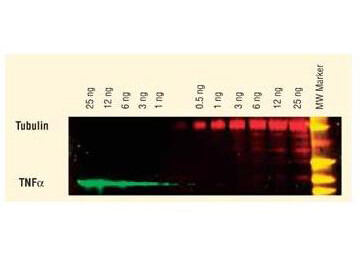
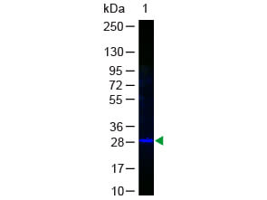
Anti-GFP Biotin Conjugated Antibody has been tested by ELISA and western blot and is suitable for immunoblotting, ELISA, immunohistochemistry, immunomicroscopy as well as other antibody based assays using streptavidin or avidin conjugates requiring lot-to-lot consistency.
Formulation
1.0 mg/mL by UV absorbance at 280 nm
0.02 M Potassium Phosphate, 0.15 M Sodium Chloride, pH 7.2
0.01% (w/v) Sodium Azide
10 mg/mL Bovine Serum Albumin (BSA) - Immunoglobulin and Protease free
1.0 mL
Restore with deionized water (or equivalent)
Shipping & Handling
Ambient
Store Anti-GFP at 4° C prior to restoration. For extended storage aliquot contents and freeze at -20° C or below. Avoid cycles of freezing and thawing. Centrifuge product if not completely clear after standing at room temperature. This product is stable for several weeks at 4° C as an undiluted liquid. Dilute only prior to immediate use.
Expiration date is one (1) year from date of receipt.
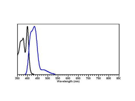
Conjugated Anti-GFP is ideal for western blotting, ELISA and Immunohistochemistry. Green fluorescent protein is a 27 kDa protein produced from the jellyfish Aequorea victoria, which emits green light (emission peak at a wavelength of 509nm) when excited by blue light. GFP is an important tool in cell biology research. GFP is widely used enabling researchers to visualize and localize GFP-tagged proteins within living cells without the need for chemical staining.
Daw TB et al. (2023). Direct Comparison of Epifluorescence and Immunostaining for Assessing Viral Mediated Gene Expression in the Primate Brain. Hum Gene Ther.
Applications
IF, Confocal Microscopy; IHC, ICC, Histology
Lu H et al. (2023). Alternative splicing and heparan sulfation converge on neurexin-1 to control glutamatergic transmission and autism-related behaviors. Cell Rep.
Applications
Bead Conjugation
Kim YE et al. (2023). Reversibility and developmental neuropathology of linear nevus sebaceous syndrome caused by dysregulation of the RAS pathway. Cell Rep.
Applications
IF, Confocal Microscopy
Stajano D et al. (2023). Tetraspanin depletion impairs extracellular vesicle docking at target neurons. ISEV journals
Applications
Immuno-gold Electron Microscopy
Jones, T et al. (2022). Differential requirements for the Eps15 homology domain proteins EHD4 and EHD2 in the regulation of mammalian ciliogenesis. Traffic (Copenhagen, Denmark)
Applications
IF, Confocal Microscopy
De Mazière A et al. (2022). An optimized protocol for immuno-electron microscopy of endogenous LC3. Autophagy.
Applications
Immuno-gold Electron Microscopy
Oorschot V et al. (2021). TEM, SEM, and STEM-based immuno-CLEM workflows offer complementary advantages. Sci Rep.
Applications
IF, Confocal Microscopy; TEM
Li H et al. (2021). Cellular requirements for PIN polar cargo clustering in Arabidopsis thaliana. New Phytol.
Applications
Immuno-EM
Zulkefli K et al. (2021). A role for Rab30 in retrograde trafficking and maintenance of endosome-TGN organization. Exp Cell Res.
Applications
Undefined
Wieghofer P et al. (2021). Mapping the origin and fate of myeloid cells in distinct compartments of the eye by single‐cell profiling. EMBO J.
Applications
IF, Confocal Microscopy
Tran NH et al. (2021). The stress-sensing domain of activated IRE1α forms helical filaments in narrow ER membrane tubes. Science.
Applications
Immuno-EM
Jongsma ML et al. (2020). SKIP-HOPS recruits TBC1D15 for a Rab7-to-Arl8b identity switch to control late endosome transport. EMBO J.
Applications
Immuno-EM
Cushnie AK et al. (2020). Using rAAV2-retro in rhesus macaques: promise and caveats for circuit manipulation. J Neurosci Methods.
Applications
IHC, ICC, Histology
Bohlen MO et al. (2020). Adeno-Associated Virus Capsid-Promoter Interactions in the Brain Translate from Rat to the Nonhuman Primate. Hum Gene Ther.
Applications
IF, Confocal Microscopy
Cabukusta B et al. (2020). Human VAPome Analysis Reveals MOSPD1 and MOSPD3 as Membrane Contact Site Proteins Interacting with FFAT-Related FFNT Motifs. Cell Rep.
Applications
WB, IB, PCA
Bohlen MO, El-Nahal HG, Sommer MA. (2019). Transduction of Craniofacial Motoneurons Following Intramuscular Injections of Canine Adenovirus Type-2 (CAV-2) in Rhesus Macaques. Front Neuroanat.
Applications
IF, Confocal Microscopy
Andres-Alonso M, Ammar MR, Butnaru I, et al. (2019). SIPA1L2 controls trafficking and local signaling of TrkB-containing amphisomes at presynaptic terminals. Nat Commun.
Applications
Immuno-EM
Croop B et al. (2019). Facile single-molecule pull-down assay for analysis of endogenous proteins. Phys Biol.
Applications
SiMPull
Yousefi OS et al. (2019). Optogenetic control shows that kinetic proofreading regulates the activity of the T cell receptor. Elife.
Applications
FC, FACS, FLOW
Lee JY et al. (2019). Limiting Neuronal Nogo Receptor 1 Signaling during Experimental Autoimmune Encephalomyelitis Preserves Axonal Transport and Abrogates Inflammatory Demyelination. J Neurosci.
Applications
Immuno-EM
Jangphattananont N et al. (2019). Distinct localization of mature HGF from its precursor form in developing and repairing the stomach. Int J Mol Sci.
Applications
IHC, ICC, Histology
Koerver L et al. (2019). The ubiquitin‐conjugating enzyme UBE 2 QL 1 coordinates lysophagy in response to endolysosomal damage. EMBO Rep.
Applications
Immuno-EM
Zöller et al. (2018). Silencing of TGFβ signalling in microglia results in impaired homeostasis. Nature Communications
Applications
IF, Confocal Microscopy
Liebmann et al. (2018). Regulation of Neuronal Na,K-ATPase by Extracellular Scaffolding Proteins. International Journal of Molecular Sciences
Applications
Quantum Dot Labeling (Q-Dot)
Maeder CI, Kim JI, Liang X, et al. (2018). The THO Complex Coordinates Transcripts for Synapse Development and Dopamine Neuron Survival. Cell.
Applications
IP, Co-IP
Ge Y et al. (2018). Clptm1 limits forward trafficking of GABAA receptors to scale inhibitory synaptic strength. Neuron.
Applications
Ab Purification
McArthur K et al. (2018). BAK/BAX macropores facilitate mitochondrial herniation and mtDNA efflux during apoptosis. Science.
Applications
Immuno-EM
Bender J et al. (2018). Multiplexed antibody detection from blood sera by immobilization of in vitro expressed antigens and label-free readout via imaging reflectometric interferometry (iRIf). Biosens Bioelectron.
Applications
Other
Bower NI et al. (2017). Mural lymphatic endothelial cells regulate meningeal angiogenesis in the zebrafish. Nat Neurosci.
Applications
Immuno-EM
Goldmann et al. (2016). Origin, fate and dynamics of macrophages at central nervous system interfaces. Nature Immunology
Applications
IF, Confocal Microscopy; Multiplex Assay
Sztal et al. (2015). Zebrafish models for nemaline myopathy reveal a spectrum of nemaline bodies contributing to reduced muscle function. Acta Neuropathologica
Applications
IHC, ICC, Histology
Baek et al. (2015). An AKT3-FOXG1-reelin network underlies defective migration in human focal malformations of cortical development. Nature Medicine
Applications
IF, Confocal Microscopy; IHC, ICC, Histology; Multiplex Assay
Henau et al. (2015). A redox signalling globin is essential for reproduction in Caenorhabditis elegans. Nature Communications
Applications
TEM
Oorschot VMJ et al. (2014). Immuno correlative light and electron microscopy on Tokuyasu cryosections. Methods Cell Biol.
Applications
IF, Confocal Microscopy; Immuno-EM
Kang Y et al. (2014). A combined transgenic proteomic analysis and regulated trafficking of neuroligin-2. J Biol Chem.
Applications
Ab Purification
Goldmann T et al. (2013). A new type of microglia gene targeting shows TAK1 to be pivotal in CNS autoimmune inflammation. Nat Neurosci.
Applications
IF, Confocal Microscopy
Kunte, A et al. (2013). Endoplasmic reticulum glycoprotein quality control regulates CD1d assembly and CD1d-mediated antigen presentation. The Journal of Biological Chemistry
Applications
WB, IB, PCA
Jain A et al. (2012). Single-molecule pull-down for studying protein interactions. Nat Protoc.
Applications
SiMPull
Panter MS et al. (2012). Dynamics of major histocompatibility complex class I association with the human peptide-loading complex. J Biol Chem.
Applications
IF, Confocal Microscopy
Jain, A et al. (2011). Probing cellular protein complexes using single-molecule pull-down. Nature
Applications
WB, IB, PCA
Graf ER et al. (2004). Neurexins induce differentiation of GABA and glutamate postsynaptic specializations via neuroligins. Cell.
Applications
Ab Immobilization
This product is for research use only and is not intended for therapeutic or diagnostic applications. Please contact a technical service representative for more information. All products of animal origin manufactured by Rockland Immunochemicals are derived from starting materials of North American origin. Collection was performed in United States Department of Agriculture (USDA) inspected facilities and all materials have been inspected and certified to be free of disease and suitable for exportation. All properties listed are typical characteristics and are not specifications. All suggestions and data are offered in good faith but without guarantee as conditions and methods of use of our products are beyond our control. All claims must be made within 30 days following the date of delivery. The prospective user must determine the suitability of our materials before adopting them on a commercial scale. Suggested uses of our products are not recommendations to use our products in violation of any patent or as a license under any patent of Rockland Immunochemicals, Inc. If you require a commercial license to use this material and do not have one, then return this material, unopened to: Rockland Inc., P.O. BOX 5199, Limerick, Pennsylvania, USA.


