GFP Antibody Peroxidase Conjugate
Goat Polyclonal
25 References
600-103-215
1 mg
Lyophilized
WB, ELISA, IHC, IF, IP, Multiplex
GFP, eGFP, rGFP
Goat
Shipping info:
$50.00 to US & $70.00 to Canada for most products. Final costs are calculated at checkout.
Product Details
Anti-GFP (GOAT) Antibody Peroxidase Conjugated Min X Hu Ms and Rt Serum Proteins - 600-103-215
goat anti-GFP Antibody peroxidase Conjugation, HRP conjugated goat anti-GFP antibody, Green Fluorescent Protein, GFP antibody, Green Fluorescent Protein antibody, EGFP, enhanced Green Fluorescent Protein, Aequorea victoria, Jellyfish
Goat
Peroxidase (HRP)
Polyclonal
IgG
Target Details
GFP, eGFP, rGFP
Recombinant Protein
Recombinant Green Fluorescent Protein (GFP) fusion protein corresponding to the full length amino acid sequence (246aa) derived from the jellyfish Aequorea victoria.
Anti-GFP antibody was prepared from monospecific antiserum by immunoaffinity chromatography using Green Fluorescent Protein (Aequorea victoria) coupled to agarose beads followed by solid phase adsorption(s) to remove any unwanted reactivities. Assay by immunoelectrophoresis resulted in a single precipitin arc against anti-Goat Serum, anti-Peroxidase and purified and partially purified Green Fluorescent Protein (Aequorea victoria). No reaction was observed against Human, Mouse and Rat Serum Proteins.
P42212 - UniProtKB
Application Details
ELISA, WB
IF, IHC, IP, Multiplex
- View References
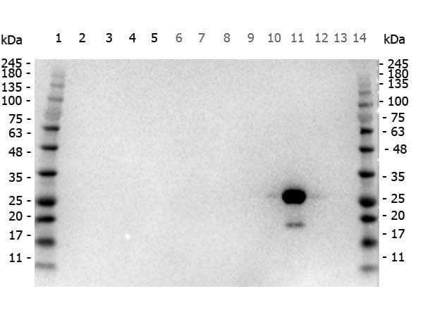
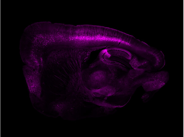
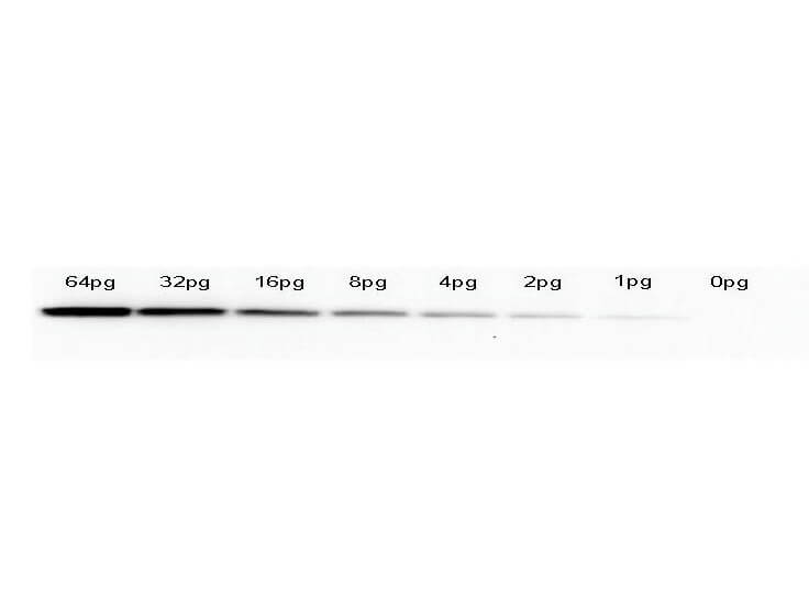
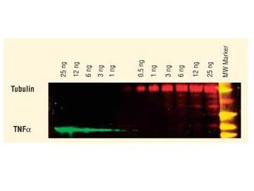
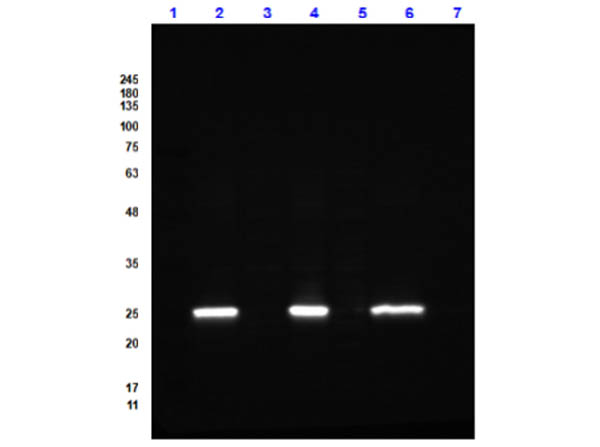
Polyclonal anti-GFP is designed to detect GFP and its variants. Anti-GFP Peroxidase conjugated antibody has been tested by ELISA to detect GFP by ELISA (sandwich or capture) for the direct binding of antigen and recognizes wild type, recombinant and enhanced forms of GFP and by western blot. Biotin conjugated polyclonal anti-GFP used in a sandwich ELISA is well suited to titrate GFP in solution using this antibody in combination with Rockland's monoclonal anti-GFP (600-301-215) using either form of the antibody as the capture or detection antibody. However, use the monoclonal form only for the detection of wild type or recombinant GFP as this form does not sufficiently detect 'enhanced' GFP. The detection antibody is typically conjugated to biotin and subsequently reacted with streptavidin conjugated HRP (code # S000-03). Fluorochrome conjugated polyclonal anti-GFP can be used to detect GFP by immunofluorescence microscopy in prokaryotic (E.coli) and eukaryotic (CHO cells) expression systems and can detect GFP containing inserts. Significant amplification of signal is achieved using fluorochrome conjugated polyclonal anti-GFP relative to the fluorescence of GFP alone. For immunoblotting use either alkaline phosphatase or peroxidase conjugated polyclonal anti-GFP to detect GFP or GFP containing proteins on western blots. Optimal titers for applications should be determined by the researcher.
Formulation
1.0 mg/mL by UV absorbance at 280 nm
0.02 M Potassium Phosphate, 0.15 M Sodium Chloride, pH 7.2
0.01% (w/v) Gentamicin Sulfate. Do NOT add Sodium Azide!
10 mg/mL Bovine Serum Albumin (BSA) - Immunoglobulin and Protease free
1.0 mL
Restore with deionized water (or equivalent)
Shipping & Handling
Ambient
Store GFP antibody at 4° C prior to restoration. For extended storage aliquot contents and freeze at -20° C or below. Avoid cycles of freezing and thawing. Centrifuge product if not completely clear after standing at room temperature. This product is stable for several weeks at 4° C as an undiluted liquid. Dilute only prior to immediate use.
Expiration date is one (1) year from date of receipt.
HRP Anti-GFP is ideal for western blotting, ELISA and Immunohistochemistry. Green fluorescent protein is a 27 kDa protein produced from the jellyfish Aequorea victoria, which emits green light (emission peak at a wavelength of 509nm) when excited by blue light. GFP is an important tool in cell biology research. GFP is widely used enabling researchers to visualize and localize GFP-tagged proteins within living cells without the need for chemical staining.
Lee Y et al. (2023). Atg1-dependent phosphorylation of Vps34 is required for dynamic regulation of the phagophore assembly site and autophagy in Saccharomyces cerevisiae. Autophagy.
Applications
WB, IB, PCA
Zhang Y et al. (2022). Sex-specific characteristics of cells expressing the cannabinoid 1 receptor in the dorsal horn of the lumbar spinal cord. J Comp Neurol.
Applications
IHC, ICC, Histology
Maslakova AA et al. (2022). Towards unveiling the nature of short SERPINA1 transcripts: Avoiding the main ORF control to translate alpha1-antitrypsin C-terminal peptides. Int J Biol Macromol.
Applications
Undefined
Devireddy S et al. (2022). Efficient progranulin exit from the ER requires its interaction with prosaposin, a Surf4 cargo. J Cell Biol.
Applications
WB, IB, PCA
Angarola B et al. (2021). LyTS: A Lysosome Localized Complex of TMEM192 and STK11IP. bioRxiv Preprint
Applications
WB, IB, PCA
Devireddy S et al. (2021). Surf4 Promotes Endoplasmic Reticulum Exit of the Lysosomal Prosaposin-Progranulin Complex. bioRxiv Preprint
Applications
WB, IB, PCA
Stepanik V et al. (2020). FGF Pyramus Has a Transmembrane Domain and Cell-Autonomous Function in Polarity. Curr Biol.
Applications
IF, Confocal Microscopy
Lim G et al. (2020). Phosphoregulation of Rad51/Rad52 by CDK1 functions as a molecular switch for cell cycle–specific activation of homologous recombination. Sci Adv.
Applications
WB, IB, PCA
Morshed S et al. (2020). TORC1 regulates ESCRT-0 complex formation on the vacuolar membrane and microautophagy induction in yeast. Biochem Biophys Res Commun.
Applications
WB, IB, PCA
Del Rosario JS et al. (2020). Gi‐coupled receptor activation potentiates Piezo2 currents via Gβγ. EMBO Rep.
Applications
WB, IB, PCA
Hennigan RF et al. (2019). Proximity biotinylation identifies a set of conformation-specific interactions between Merlin and cell junction proteins. Sci Signal.
Applications
WB, IB, PCA
Kim Y et al. (2019). Global analysis of protein homomerization in Saccharomyces cerevisiae. Genome Res.
Applications
WB, IB, PCA
Macabenta F et al. (2019). Migrating cells control morphogenesis of substratum serving as track to promote directional movement of the collective. Development.
Applications
IF, Confocal Microscopy
Atabekova AK et al. (2018). Mechanical stress-induced subcellular re-localization of N-terminally truncated tobacco Nt-4/1 protein. Biochimie.
Applications
WB, IB, PCA
Sun J et al. (2018). FGF controls epithelial-mesenchymal transitions during gastrulation by regulating cell division and apicobasal polarity. Development.
Applications
IF, Confocal Microscopy; Multiplex Assay
Bisgrove et al. (2017). Maternal Gdf3 is an obligatory cofactor in Nodal signaling for embryonic axis formation in zebrafish. Elife
Applications
IHC, ICC, Histology
Gustin et al. (2015). Ebola Virus Glycoprotein Promotes Enhanced Viral Egress by Preventing Ebola VP40 From Associating With the Host Restriction Factor BST2/Tetherin. The Journal of Infectious Diseases
Applications
IP, Co-IP; WB, IB, PCA
Fahrenkamp et al. (2015). Src family kinases interfere with dimerization of STAT5A through a phosphotyrosine-SH2 domain interaction. Cell Communication and Signaling
Applications
IP, Co-IP
Botto et al. (2015). Kaposi Sarcoma Herpesvirus Induces HO-1 during De Novo Infection of Endothelial Cells via Viral miRNA-Dependent and -Independent Mechanisms. mBio
Applications
WB, IB, PCA
Bejarano E et al. (2014). Connexins modulate autophagosome biogenesis. Nat Cell Biol.
Applications
WB, IB, PCA
Sung MK et al. (2013). Genome-wide bimolecular fluorescence complementation analysis of SUMO interactome in yeast. Genome Res.
Applications
IP, Co-IP; WB, IB, PCA
Chatain N et al. (2013). Src family kinases mediate cytoplasmic retention of activated STAT5 in BCR–ABL-positive cells. Oncogene.
Applications
IP, Co-IP
Ha CW et al. (2012). Nsi1 plays a significant role in the silencing of ribosomal DNA in Saccharomyces cerevisiae. Nucleic Acids Res.
Applications
WB, IB, PCA
Guarino E et al. (2011). Cdt1 proteolysis is promoted by dual PIP degrons and is modulated by PCNA ubiquitylation. Nucleic Acids Res.
Applications
WB, IB, PCA
Sugita M et al. (2004). Molecular dissection of the butyrate action revealed the involvement of mitogen-activated protein kinase in cystic fibrosis transmembrane conductance regulator biogenesis. Mol Pharmacol.
Applications
IF, Confocal Microscopy; WB, IB, PCA
This product is for research use only and is not intended for therapeutic or diagnostic applications. Please contact a technical service representative for more information. All products of animal origin manufactured by Rockland Immunochemicals are derived from starting materials of North American origin. Collection was performed in United States Department of Agriculture (USDA) inspected facilities and all materials have been inspected and certified to be free of disease and suitable for exportation. All properties listed are typical characteristics and are not specifications. All suggestions and data are offered in good faith but without guarantee as conditions and methods of use of our products are beyond our control. All claims must be made within 30 days following the date of delivery. The prospective user must determine the suitability of our materials before adopting them on a commercial scale. Suggested uses of our products are not recommendations to use our products in violation of any patent or as a license under any patent of Rockland Immunochemicals, Inc. If you require a commercial license to use this material and do not have one, then return this material, unopened to: Rockland Inc., P.O. BOX 5199, Limerick, Pennsylvania, USA.

