Datasheet is currently unavailable. Try again or CONTACT US
GFP Antibody Fluorescein Conjugated
Mouse Monoclonal 9F9.F9 IgG1 kappa
4 References
600-302-215
1 mg
Lyophilized
WB, IHC, IF, FC, Dot Blot, Multiplex
GFP, eGFP, rGFP
Mouse
Shipping info:
$50.00 to US & $70.00 to Canada for most products. Final costs are calculated at checkout.
Product Details
Anti-GFP (MOUSE) Monoclonal Antibody Fluorescein Conjugated - 600-302-215
mouse anti-GFP antibody FITC conjugation, fluorescein conjugated mouse anti-GFP antibody, Green Fluorescent Protein, GFP antibody, Green Fluorescent Protein antibody, EGFP, enhanced Green Fluorescent Protein, Aequorea victoria, Jellyfish
Mouse
Fluorescein (FITC)
Monoclonal
IgG
Target Details
GFP, eGFP, rGFP
Recombinant Protein
Anti-Green Fluorescent Protein (GFP) is produced by immunizing GFP containing fusion protein that corresponds to the full length amino acid sequence (246aa) derived from the jellyfish Aequorea victoria.
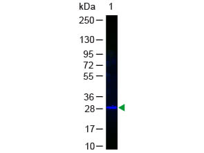
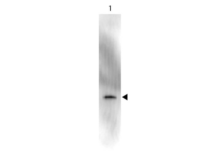
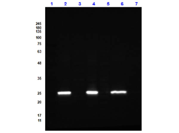
GFP Fluorescein Conjugated Antibody was prepared from tissue culture supernatant by Protein A affinity chromatography. Assay by Immunoelectrophoresis resulted in a single precipitin arc against anti-fluorescein and anti-Mouse Serum. Reactivity is observed against recombinant Green Fluorescent Protein (000-001-215, recombinant GFP from Aequorea victoria) by Western blot. No reaction is seen against RFP.
P42212 - UniProtKB
Application Details
Dot Blot, WB
FC, IF, IHC, Multiplex
- View References
Monoclonal anti-GFP is designed to detect enhanced GFP and GFP containing recombinant proteins. This antibody can be used to detect GFP by ELISA (sandwich or capture) for the direct binding of antigen. Biotin conjugated monoclonal anti-GFP is well suited to titrate GFP in a sandwich ELISA in combination with Rockland's polyclonal anti-GFP (600-101-215) as the capture antibody. Only use the monoclonal form for the detection of enhanced or recombinant GFP. Polyclonal anti-GFP detects all variants of GFP tested to date. The biotin conjugated detection antibody is typically used with streptavidin conjugated HRP (code # S000-03) or other streptavidin conjugates. The use of polyclonal anti-GFP results in significant amplification of signal when fluorochrome conjugated polyclonal anti-GFP is used relative to the fluorescence of GFP alone. This antibody was tested by western blotting, for immunoblotting use either alkaline phosphatase or peroxidase conjugated anti-GFP to detect GFP or GFP containing proteins on western blots. Optimal titers for applications should be determined by the researcher.
Formulation
0.02 M Potassium Phosphate, 0.15 M Sodium Chloride, pH 7.2
0.01% (w/v) Sodium Azide
10 mg/mL Bovine Serum Albumin (BSA) - Immunoglobulin and Protease free
1.0 mL
Restore with deionized water (or equivalent)
Shipping & Handling
Ambient
Store vial at 4° C prior to restoration. For extended storage aliquot contents and freeze at -20° C or below. Avoid cycles of freezing and thawing. Centrifuge product if not completely clear after standing at room temperature. This product is stable for several weeks at 4° C as an undiluted liquid. Dilute only prior to immediate use.
Expiration date is one (1) year from date of receipt.
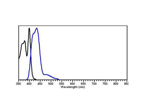
Green fluorescent protein is a 27 kDa protein produced from the jellyfish Aequorea victoria, which emits green light (emission peak at a wavelength of 509nm) when excited by blue light. GFP is an important tool in cell biology research. GFP is widely used enabling researchers to visualize and localize GFP-tagged proteins within living cells without the need for chemical staining.
Godfrey RK et al. (2023). Modelling TDP-43 proteinopathy in Drosophila uncovers shared and neuron-specific targets across ALS and FTD relevant circuits. Acta Neuropathol Commun.
Applications
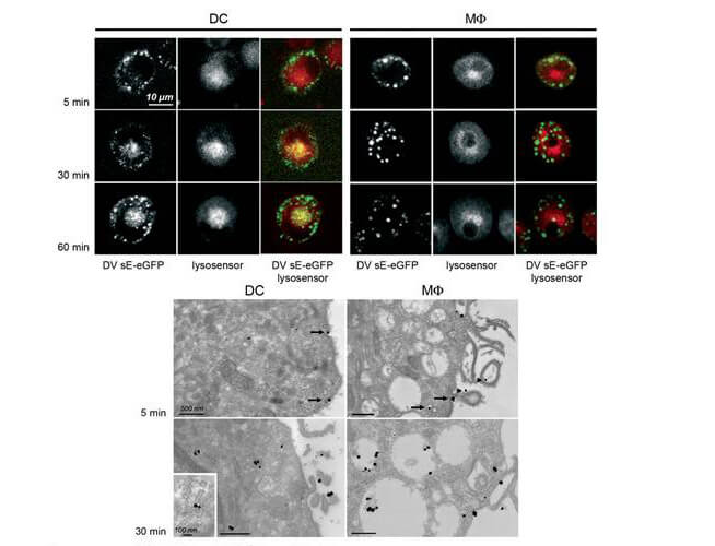
IF, Confocal Microscopy
Collins, CM et al. (2012). Tracking murine gammaherpesvirus 68 infection of germinal center B cells in vivo. PloS One
Applications
IF, Confocal Microscopy; Multiplex Assay
Marks BR et al. (2009). Thymic self-reactivity selects natural interleukin 17-producing T cells that can regulate peripheral inflammation. Nat Immunol.
Applications
FC, FACS, FLOW; Multiplex Assay
Athena M Soulika et al. (2009). Initiation and progression of axonopathy in experimental autoimmune encephalomyelitis. J Neurosci.
Applications
IHC, ICC, Histology; Multiplex Assay
This product is for research use only and is not intended for therapeutic or diagnostic applications. Please contact a technical service representative for more information. All products of animal origin manufactured by Rockland Immunochemicals are derived from starting materials of North American origin. Collection was performed in United States Department of Agriculture (USDA) inspected facilities and all materials have been inspected and certified to be free of disease and suitable for exportation. All properties listed are typical characteristics and are not specifications. All suggestions and data are offered in good faith but without guarantee as conditions and methods of use of our products are beyond our control. All claims must be made within 30 days following the date of delivery. The prospective user must determine the suitability of our materials before adopting them on a commercial scale. Suggested uses of our products are not recommendations to use our products in violation of any patent or as a license under any patent of Rockland Immunochemicals, Inc. If you require a commercial license to use this material and do not have one, then return this material, unopened to: Rockland Inc., P.O. BOX 5199, Limerick, Pennsylvania, USA.

