GFP Antibody
Chicken Polyclonal
39 References
600-901-215S
600-901-215
25 µL
100 µg
Liquid (sterile filtered)
Liquid (sterile filtered)
WB, ELISA, IHC, IF, Dot Blot, EM, IP, Multiplex, Purification
GFP, eGFP, rGFP
Chicken
Shipping info:
$50.00 to US & $70.00 to Canada for most products. Final costs are calculated at checkout.
Product Details
Anti-GFP (CHICKEN) Antibody - 600-901-215
chicken anti-GFP antibody, Green Fluorescent Protein, GFP antibody, Green Fluorescent Protein antibody, EGFP, enhanced Green Fluorescent Protein, Aequorea victoria, Jellyfish
Chicken
Polyclonal
IgY
Target Details
GFP, eGFP, rGFP
Recombinant Protein
The immunogen is a Green Fluorescent Protein (GFP) fusion protein corresponding to the full length amino acid sequence (246aa) derived from the jellyfish Aequorea victoria.
Anti-GFP IgY was prepared from egg yolks by immunoaffinity chromatography using Green Fluorescent Protein (Aequorea victoria) coupled to agarose beads followed by solid phase adsorption(s) to remove any unwanted reactivities. Assay by immunoelectrophoresis resulted in a single precipitin arc against anti-Chicken Serum and purified and partially purified Green Fluorescent Protein (Aequorea victoria). No reaction was observed against Human, Mouse or Rat serum proteins.
Application Details
Dot Blot, ELISA, WB
EM, IF, IHC, IP, Purification, Multiplex
- View References
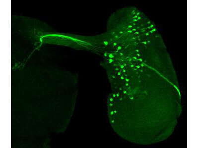
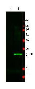
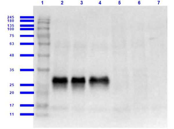
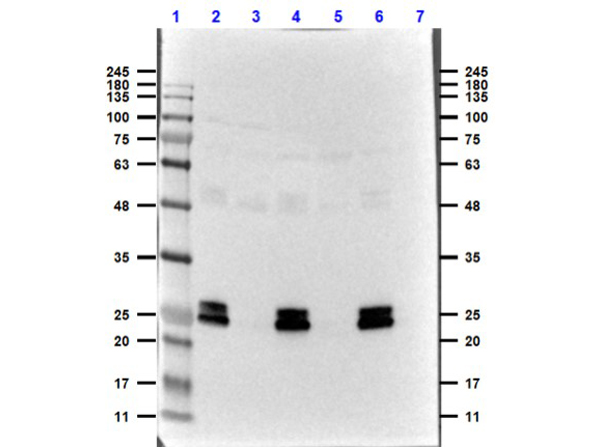
Polyclonal anti-GFP is designed to detect GFP and its variants. Anti-GFP Antibody has been tested by ELISA, dot blot, and western and is suitable in immunofluorescence. This antibody can be used to detect GFP by ELISA (sandwich or capture) for the direct binding of antigen and recognizes wild type, recombinant and enhanced forms of GFP. Biotin conjugated polyclonal anti-GFP used in a sandwich ELISA is well suited to titrate GFP in solution using this antibody in combination with Rockland´s monoclonal anti-GFP (600-301-215) using either form of the antibody as the capture or detection antibodies. However, use the monoclonal form only for the detection of wild type or recombinant GFP as this form does not sufficiently detect ´enhanced´ GFP. The detection antibody is typically conjugated to biotin and subsequently reacted with streptavidin conjugated HRP (code # S000-03). Fluorochrome conjugated polyclonal anti-GFP can be used to detect GFP by immunofluorescence microscopy in prokaryotic (E.coli) and eukaryotic (CHO cells) expression systems and can detect GFP containing inserts. Significant amplification of signal is achieved using fluorochrome conjugated polyclonal anti-GFP relative to the fluorescence of GFP alone. For immunoblotting use either alkaline phosphatase or peroxidase conjugated polyclonal anti-GFP to detect GFP or GFP containing proteins on western blots.
Formulation
1.18 mg/ml by UV absorbance at 280 nm
0.02 M Potassium Phosphate, 0.15 M Sodium Chloride, pH 7.2
0.01% (w/v) Sodium Azide
None
Shipping & Handling
Dry Ice
Store GFP Antibody at -20° C prior to opening. Aliquot contents and freeze at -20° C or below for extended storage. Avoid cycles of freezing and thawing. Centrifuge product if not completely clear after standing at room temperature. This product is stable for several weeks at 4° C as an undiluted liquid. Dilute only prior to immediate use.
Expiration date is one (1) year from date of receipt.
Anti-GFP antibody is designed for immunofluorescence microscopy, fluorescence based plate assays (FLISA) and fluorescent western blotting. GFP antibody is also suitable for multiplex analysis, including multicolor imaging, utilizing various commercial platforms. Chicken IgY lacks the classic "Fc" domain and does not bind to mammalian IgG Fc receptors. Thus resulting in lower backgrounds for western blotting, ELISA and Immunohistochemistry.
Xing YL et al. (2025). BRAF/MEK Inhibition Induces Cell State Transitions Boosting Immune Checkpoint Sensitivity in BRAFV600E -mutant Glioma. Cell Rep Med.
Applications
IF, Confocal Microscopy
Lin CY et al. (2025). Sigma-1R-Pom121 axis preserves nuclear transport and integrity in poly-PR-induced C9orf72 ALS. Neurobiol Dis.
Applications
IF, Confocal Microscopy; WB, IB, PCA
Sakamoto H et al. (2024). Membrane-Targeted palGFP Predominantly Localizes to the Plasma Membrane but not to Neurosecretory Vesicle Membranes in Rat Oxytocin Neurons. Acta Histochem Cytochem.
Applications
IF, Confocal Microscopy
Pires PM et al. (2024). Converting an allocentric goal into an egocentric steering signal. Nature.
Applications
IHC, ICC, Histology
Vijayan V et al. (2023). A rise-to-threshold process for a relative-value decision. Nature.
Applications
IF, Confocal Microscopy
Roy S et al. (2023). Large-scale interrogation of retinal cell functions by 1-photon light-sheet microscopy. Cell Rep Methods.
Applications
IF, Confocal Microscopy
Phan QM et al. (2023). Lineage commitment of dermal fibroblast progenitors is controlled by Kdm6b-mediated chromatin demethylation. EMBO J.
Applications
IF, Confocal Microscopy
Ciesielski HM et al. (2022). Erebosis, a new cell death mechanism during homeostatic turnover of gut enterocytes. PLoS Biol.
Applications
Immuno-EM
Lamontagne, JO et al. (2022). Transcription factors AP-2α and AP-2β regulate distinct segments of the distal nephron in the mammalian kidney. Nature Communications
Applications
IF, Confocal Microscopy
Lyu C et al. (2022). Building an allocentric travelling direction signal via vector computation. Nature.
Applications
IHC, ICC, Histology
Zhao X et al. (2022). 4D imaging analysis of the aging mouse neural stem cell niche reveals a dramatic loss of progenitor cell dynamism regulated by the RHO-ROCK pathway. Stem Cell Reports.
Applications
IHC, ICC, Histology
Groschner LN et al. (2022). A biophysical account of multiplication by a single neuron. Nature
Applications
IHC, ICC, Histology
Fenk LM et al. (2022). Muscles that move the retina augment compound eye vision in Drosophila. Nature.
Applications
IF, Confocal Microscopy
Oti T et al. (2021). Oxytocin influences male sexual activity via non-synaptic axonal release in the spinal cord. Curr Biol.
Applications
IHC, ICC, Histology
Li, Y et al. (2021). Genetic analysis of the septal peptidoglycan synthase FtsWI complex supports a conserved activation mechanism for SEDS-bPBP complexes. PloS Genetics
Applications
WB, IB, PCA
Wang, Q et al. (2021). The Extracellular Superoxide Dismutase Sod5 From Fusarium oxysporum Is Localized in Response to External Stimuli and Contributes to Fungal Pathogenicity. Frontiers in Plant Science
Applications
WB, IB, PCA
Osso LA et al. (2021). Experience-dependent myelination following stress is mediated by the neuropeptide dynorphin. Neuron.
Applications
IHC, ICC, Histology
Nam HS, Capecchi MR. (2020). Lrig1 expression prospectively identifies stem cells in the ventricular-subventricular zone that are neurogenic throughout adult life. Neural Dev.
Applications
IF, Confocal Microscopy
Wu et al. (2020). let-7-Complex MicroRNAs Regulate Broad-Z3, Which Together with Chinmo Maintains Adult Lineage Neurons in an Immature State. G3 Genes Genomics Genetics
Applications
IF, Confocal Microscopy
Sanders DW et al. (2020). Competing protein-RNA interaction networks control multiphase intracellular organization. Cell.
Applications
IF, Confocal Microscopy; WB, IB, PCA
Duan ZRS et al. (2020). GABAergic restriction of network dynamics regulates interneuron survival in the developing cortex. Neuron.
Applications
IHC, ICC, Histology
Goswami et al. (2019). Novel window for cancer nanotheranostics: non-invasive ocular assessments of tumor growth and nanotherapeutic treatment efficacy in vivo. Biomedical Optics Express
Applications
IHC, ICC, Histology
Brito, C et al. (2019). Perfringolysin O-Induced Plasma Membrane Pores Trigger Actomyosin Remodeling and Endoplasmic Reticulum Redistribution. Toxins
Applications
TEM
Liu, W et al. (2019). Alp7-Mto1 and Alp14 synergize to promote interphase microtubule regrowth from the nuclear envelope. Journal of Molecular Cell Biology
Applications
IP, Co-IP; WB, IB, PCA
Chiu et al. (2018). Input-Specific NMDAR-Dependent Potentiation of Dendritic GABAergic Inhibition. Neuron
Applications
IF, Confocal Microscopy
Li et al. (2018). Hub-organized parallel circuits of central circadian pacemaker neurons for visual photoentrainment in Drosophila. Nature Communications
Applications
IF, Confocal Microscopy
Fujiwara Y et al. (2018). The CCHamide1 Neuropeptide Expressed in the Anterior Dorsal Neuron 1 Conveys a Circadian Signal to the Ventral Lateral Neurons in Drosophila melanogaster. Front Physiol.
Applications
IHC, ICC, Histology
Kronenberg G et al. (2018). Distinguishing features of microglia-and monocyte-derived macrophages after stroke. Acta Neuropathol.
Applications
IHC, ICC, Histology
Oti T et al. (2018). Effects of sex steroids on the spinal gastrin-releasing peptide system controlling male sexual function in rats. Endocrinology.
Applications
IF, Confocal Microscopy; WB, IB, PCA
Ribeiro IMA, Drews M, Bahl A, Machacek C, Borst A, Dickson BJ. (2018). Visual Projection Neurons Mediating Directed Courtship in Drosophila. Cell.
Applications
IF, Confocal Microscopy
Maruoka et al. (2017). Nectin-4 co-stimulates the prolactin receptor by interacting with SOCS1 and inhibiting its activity on the JAK2-STAT5a signaling pathway. Journal of Biological Chemistry
Applications
WB, IB, PCA
Neukomm LJ et al. (2017). Axon death pathways converge on axundead to promote functional and structural axon disassembly. Neuron.
Applications
IF, Confocal Microscopy
Kim, AJ et al. (2017). Quantitative Predictions Orchestrate Visual Signaling in Drosophila. Cell
Applications
Electrophysiology; IHC, ICC, Histology
Boye et al. (2016). Highly Efficient Delivery of Adeno-Associated Viral Vectors to the Primate Retina. Human Gene Therapy
Applications
IHC, ICC, Histology; Multiplex Assay
Chen et al. (2015). Lin-28 promotes symmetric stem cell division and drives adaptive growth in the adult Drosophila intestine. Development
Applications
IHC, ICC, Histology; Multiplex Assay
Vladimir A Volkov et al. (2013). Long tethers provide high-force coupling of the Dam1 ring to shortening microtubules. Proc Natl Acad Sci U S A.
Applications
Bead Conjugation
He M. (2012). miRNA Tagging and Affinity-purification (miRAP). Bio Protoc
Applications
AB Purification; IF, Confocal Microscopy
Tseng C-Y, Firestein BL. (2011). The role of PSD-95 and cypin in morphological changes in dendrites following sublethal NMDA exposure. J. Neurosci.
Applications
IHC, ICC, Histology; Multiplex Assay
Oleinikov AV et al. (2009). High throughput functional assays of the variant antigen PfEMP1 reveal a single domain in the 3D7 Plasmodium falciparum genome that binds ICAM1 with high affinity and is targeted by naturally acquired neutralizing antibodies. PloS Pathogens
Applications
Ab Immobilization
This product is for research use only and is not intended for therapeutic or diagnostic applications. Please contact a technical service representative for more information. All products of animal origin manufactured by Rockland Immunochemicals are derived from starting materials of North American origin. Collection was performed in United States Department of Agriculture (USDA) inspected facilities and all materials have been inspected and certified to be free of disease and suitable for exportation. All properties listed are typical characteristics and are not specifications. All suggestions and data are offered in good faith but without guarantee as conditions and methods of use of our products are beyond our control. All claims must be made within 30 days following the date of delivery. The prospective user must determine the suitability of our materials before adopting them on a commercial scale. Suggested uses of our products are not recommendations to use our products in violation of any patent or as a license under any patent of Rockland Immunochemicals, Inc. If you require a commercial license to use this material and do not have one, then return this material, unopened to: Rockland Inc., P.O. BOX 5199, Limerick, Pennsylvania, USA.


