Datasheet is currently unavailable. Try again or CONTACT US
Datasheet

Collagen I alpha 1 propeptide Antibody

Rabbit Polyclonal

4 References
600-401-D19
100 µL
Liquid
WB, ELISA, IHC, IF, FC
Human, Mouse, Rat
Rabbit
$485.00 /Per Item
Out of stock
Ships in approximately 14 days
Shipping info:

$50.00 to US & $70.00 to Canada for most products. Final costs are calculated at checkout.

Product Details

Anti-Collagen I, alpha 1 propeptide (RABBIT) Antibody - 600-401-D19
Alpha-1 type I collagen, COLA1A1
Rabbit
Polyclonal
IgG

Target Details

COL1A1  - View All COL1A1 Products
Human, Mouse, Rat
Conjugated Peptide
Anti-collagen I, alpha 1 propeptide Antibody was produced in rabbit by repeated immunizations with a synthetic peptide corresponding to amino acid residues specific to collagen 1, alpha 1 propeptide conjugated to KLH.
Anti-Collagen I alpha 1 propeptide antibody is directed against collagen I alpha 1 propeptide (C-terminal region). Anti-Collagen I alpha 1 propeptide antibody was affinity purified from monospecific antiserum by immunoaffinity purification. Anti-Collagen I alpha 1 propeptide antibody recognizes Collagen I alpha 1 propeptide from human, rat, and mouse sources. Cross reactivity with Collagen I alpha 1 propeptide from other species has not been determined. However, reactivity is also expected from most vertebrate species based on very high sequence homology.
P02452.5 - UniProtKB
NP_000079.2 - NCBI Protein

Application Details

IHC, WB
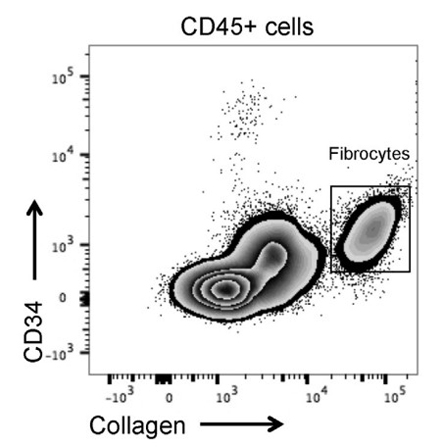
ELISA, FC, IF  - View References
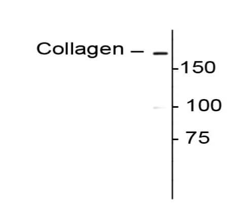
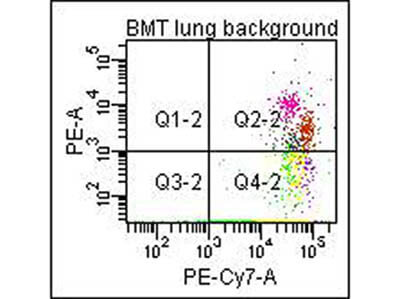
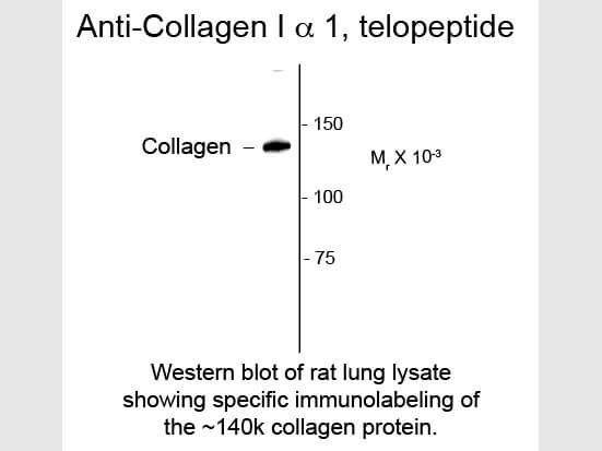
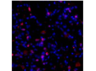
Anti-Collagen I, alpha 1 propeptide Antibody is tested for use in ELISA, Western Blotting and IHC. Specific conditions for reactivity should be optimized by the end user. Expect a band of approximately 180 kDa in size corresponding to collagen I alpha I polypeptide in human lung fibroblast extracts or other appropriate tissues or lysates. Collagen I alpha 1 propeptide antibody also works well for IHC on paraformaldehyde-fixed sections with a simple antigen-retrieval protocol (incubate slides for 20 minutes at 90º C in 10 mM sodium citrate (pH 6.0)/ 0.1 % Tween-20). Note that in paraffin sections of formaldehyde-fixed fibrotic mouse lung tissue, the antibody recognizes collagen I molecules that are still associated with the cells in which they were synthesized.

Formulation

Titrated value sufficient to run approximately 10 mini blots.
0.01 M HEPES, 0.15 M Sodium Chloride, pH 7.5
None
0.1 mg/ml Bovine Serum Albumin (BSA) - IgG and Protease free, 50% (v/v) Glycerol

Shipping & Handling

Dry Ice
Store vial at -20° C prior to opening.  This product is stable at 4° C as an undiluted liquid.  For extended storage, aliquot contents and freeze at -20° C or below.  Avoid cycles of freezing and thawing. Dilute only prior to immediate use.
Expiration date is one (1) year from date of receipt.
Collagen I alpha 1 propeptide Antibody detects collagen I which is an extracellular matrix protein that serves as a scaffold defining the shape and mechanical properties of many tissues and organs including skin, tendon, artery walls, fibrocartilage, bone and teeth. Type 1 collagen is the must abundant protein in mammals. Collagens are synthesized with N-terminal and C-terminal propeptides that are cleaved during maturation and secretion. After cleavage of the propeptides, the most N-terminal and C-terminal remaining sequences are known as telopeptides. Mutations in the collagen 1, alpha 1 gene (COL1A1) are known to cause osteogenesis imperfecta (aka brittle bone disease). Furthermore, mutations found in the fist 90 residues of the helical region of alpha 1 collagen have been implicated in the prevention or delayed removal of the procollagen N-propeptide leading to a combined osteogenesis imperfecta and Ehlers-Danlos syndrome (EDS) phenotype. Anti-Collagen I alpha 1 propeptide Antibody is ideal for investigators involved in Cell Signaling, Neuroscience, Signal Transduction research.
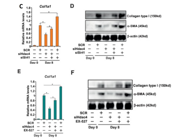
(). Sex-specific phenotypes in the aging mouse heart and consequences for chronic fibrosis. Am J Physiol Heart Circ Physiol.
Applications
FC, FACS, FLOW
(). Suppression of pancreatic ductal adenocarcinoma growth and metastasis by fibrillar collagens produced selectively by tumor cells. Nat Commun.
Applications
IHC, ICC, Histology
(). A high-content, in vitro cardiac fibrosis assay for high-throughput, phenotypic identification of compounds with anti-fibrotic activity. J Mol Cell Cardiol.
Applications
EIA, ELISA; IF, Confocal Microscopy
(). Bile acid sequestration reverses liver injury and prevents progression of nonalcoholic steatohepatitis in Western diet–fed mice. J Biol Chem.
Applications
IHC, ICC, Histology

What were the immunogens for the collagen antibodies?

The immunogens for the collagen antibodies are the respective purified solubilized native collagen proteins (tropocollagens).

In what format are the collagen antibodies provided?

The collagen antibodies are provided in liquid format at a concentration of around 1 mg/mL (Absorbance 280 nm).

In what buffer are the collagen antibodies provided?

0.02 M Potassium Phosphate, 0.15 M Sodium Chloride, pH 7.2 with 0.01% (w/v) Sodium Azide.

Are the antibodies specific to the reduced or non-reduced (native) proteins?

The collagen antibodies are specific to the native 3D epitopes found on the collagen native collagen protein. The collagen antibodies may have no reactivity to reduced collagen.

How was the reactivity of the antibodies tested? Was cross-reactivity to other collagens tested?

Each antibody was tested by immunoblot against each type of collagen (Type I – VI). Due to the close association of collagen proteins, cross-reactivity to other collagens may be observed. The optimal experimental conditions should be determined to minimize cross-reactivity in your application.

What are the recommended storage conditions for collagen antibodies?

The recommended storage temperature for collagen antibodies is -20°C. Please be sure to add glycerol 1:1 before freezing. We recommend making aliquots for best stability. The antibodies are stable for several weeks at 4°C.

Invalid lot number

If you need help finding your CoA, contact us

This product is for research use only and is not intended for therapeutic or diagnostic applications. Please contact a technical service representative for more information. All products of animal origin manufactured by Rockland Immunochemicals are derived from starting materials of North American origin. Collection was performed in United States Department of Agriculture (USDA) inspected facilities and all materials have been inspected and certified to be free of disease and suitable for exportation. All properties listed are typical characteristics and are not specifications. All suggestions and data are offered in good faith but without guarantee as conditions and methods of use of our products are beyond our control. All claims must be made within 30 days following the date of delivery. The prospective user must determine the suitability of our materials before adopting them on a commercial scale. Suggested uses of our products are not recommendations to use our products in violation of any patent or as a license under any patent of Rockland Immunochemicals, Inc. If you require a commercial license to use this material and do not have one, then return this material, unopened to: Rockland Inc., P.O. BOX 5199, Limerick, Pennsylvania, USA.