Datasheet is currently unavailable. Try again or CONTACT US
Catalase Antibody
Rabbit Polyclonal
19 References
200-4151
50 mg
Lyophilized
WB, ELISA, IHC, IF, EM, Other
Human
Rabbit
Shipping info:
$50.00 to US & $70.00 to Canada for most products. Final costs are calculated at checkout.
Product Details
Anti-Catalase (Bovine Liver) (RABBIT) Antibody (BULK ORDER) - 200-4151
rabbit anti-Catalase Antibody, Cas 1 antibody, CAT antibody, Cs1 antibody, MGC138422 antibody, MGC138424 antibody
Rabbit
Polyclonal
IgG
Target Details
CAT - View All CAT Products
Human
Native Protein
Anti-Catalase Antibody was produced by repeated immunizations with bovine liver catalase protein.
Anti-Catalase Antibody is an IgG fraction antibody purified from monospecific antiserum by a multi-step process which includes delipidation, salt fractionation and ion exchange chromatography followed by extensive dialysis against the buffer stated above. Assay by immunoelectrophoresis resulted in a single precipitin arc against anti-Rabbit Serum as well as purified and partially purified Catalase [Bovine liver]. Cross reactivity against Catalase from other tissues and species may occur but have not been specifically determined.
Application Details
ELISA, IHC, WB
EM, IF, Other
- View References
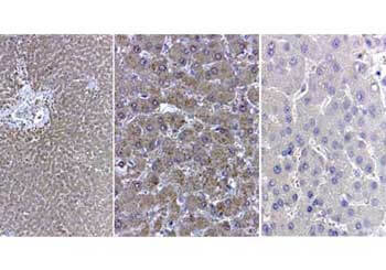
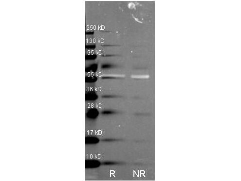
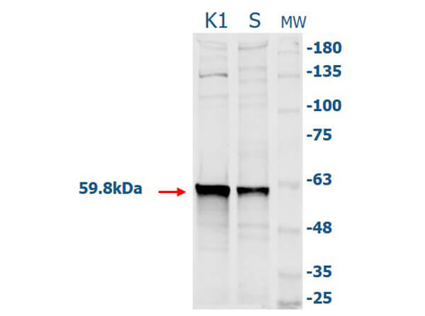
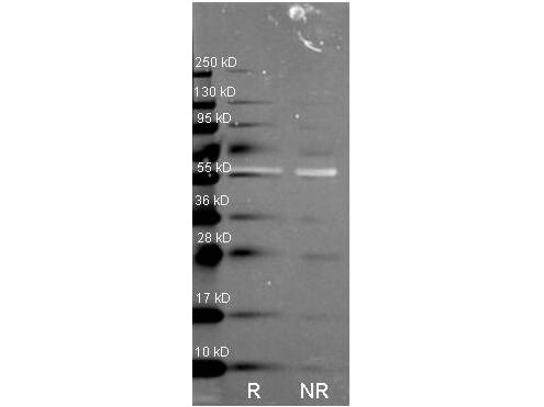
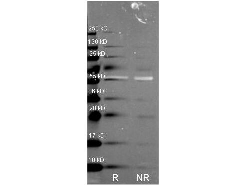
Anti-Catalase has been tested by ELISA, western blot, and immunohistochemistry. Specific conditions for reactivity should be optimized by the end user.
Formulation
10.0 mg/mL by UV absorbance at 280 nm
0.02 M Potassium Phosphate, 0.15 M Sodium Chloride, pH 7.2
0.01% (w/v) Sodium Azide
None
5.0 mL
Restore with deionized water (or equivalent)
Shipping & Handling
Ambient
Store vial at 4° C prior to restoration. For extended storage aliquot contents and freeze at -20° C or below. Avoid cycles of freezing and thawing. Centrifuge product if not completely clear after standing at room temperature. This product is stable for several weeks at 4° C as an undiluted liquid. Dilute only prior to immediate use.
Expiration date is one (1) year from date of receipt.
Anti-Catalase Antibody detects catalase. Catalase is a common enzyme found in nearly all living organisms exposed to oxygen. It catalyzes the decomposition of hydrogen peroxide to water and oxygen. Catalase is necessary in cells to protect them from the toxic effects of hydrogen peroxide and to promote growth of cells. Anti-Catalase antibody is ideal for investigators involved in Cell Signaling, Neuroscience and Signal Transduction research.
Rossi G et al. (2021). Crocetin Mitigates Irradiation Injury in an In Vitro Model of the Pubertal Testis: Focus on Biological Effects and Molecular Mechanisms. Molecules.
Applications
WB, IB, PCA
Benot-Dominguez R et al. (2021). Olive leaf extract impairs mitochondria by pro-oxidant activity in MDA-MB-231 and OVCAR-3 cancer cells. Biomed Pharmacother.
Applications
WB, IB, PCA
Morita, M et al. (2021). Generation of an immortalized astrocytic cell line from Abcd1-deficient H-2KbtsA58 mice to facilitate the study of the role of astrocytes in X-linked adrenoleukodystrophy. Heliyon
Applications
Undefined
Lehmann V et al. (2021). The potential and limitations of intrahepatic cholangiocyte organoids to study inborn errors of metabolism. J Inherit Metab Dis.
Applications
Immuno-EM
Van Veldhoven PP et al. (2020). Slc25a17 Gene Trapped Mice: PMP34 Plays a Role in the Peroxisomal Degradation of Phytanic and Pristanic Acid. Front Cell Dev Biol.
Applications
WB, IB, PCA
DiEmidio G et al. (2019). SIRT1 participates in the response to methylglyoxal-dependent glycative stress in mouse oocytes and ovary. Biochim Biophys Acta Mol Basis Dis.
Applications
WB, IB, PCA
Ruiz et al. (2017). Oxidative stress and mitochondrial dynamics malfunction are linked in Pelizaeus-Merzbacher disease. Brain Pathology
Applications
WB, IB, PCA
Kawaguchi S et al. (2013). Induction of the expressions of antioxidant enzymes by luteinizing hormone in the bovine corpus luteum. J Reprod Dev.
Applications
WB, IB, PCA
Bloch K et al. (2011). A strategy for the engineering of insulin producing cells with a broad spectrum of defense properties. Biomaterials.
Applications
WB, IB, PCA
Vincent AM, Kato K, McLean LL, Soules ME, Feldman EL. (2009). Sensory neurons and schwann cells respond to oxidative stress by increasing antioxidant defense mechanisms. Antioxid Redox Signal.
Applications
WB, IB, PCA
Jansen S et al. (2009). Glucose deprivation, oxidative stress and peroxisome proliferator-activated receptor-a (PPARA) cause peroxisome proliferation in preimplantation mouse embryos. Reproduction.
Applications
IF, Confocal Microscopy; Other; WB, IB, PCA
Fourcade S et al. (2008). Early oxidative damage underlying neurodegeneration in X-adrenoleukodystrophy. Hum Mol Genet.
Applications
WB, IB, PCA
Ma S-F et al. (2007). Combining cisplatin with cationized catalase decreases nephrotoxicity while improving antitumor activity. Kidney Int.
Applications
IHC, ICC, Histology
Takahashi H et al. (2007). High labeling indices of cdc25B is linked to progression of gastric cancers and associated with a poor prognosis. Appl Immunohistochem Mol Morphol.
Applications
IHC, ICC, Histology
Farioli-Vecchioli S et al. (2000). Inverse expression of peroxisomes and connexin-43 in the granulosa cells of the quail follicle. J Histochem Cytochem.
Applications
IF, Confocal Microscopy
Depreter M et al. (1998). Maturation of the liver-specific peroxisome versus laminin, collagen IV and integrin expression. Biol Cell.
Applications
IF, Confocal Microscopy
DeLamatre JG et al. (1996). Influence of dietary fat on the effect of endotoxin on murine hepatic peroxisomes. Hepatology.
Applications
E, EIA
Espeel M et al. (1995). Immunocytochemical localization of peroxisomal proteins in human liver and kidney. J Inherit Metab Dis.
Applications
IHC, ICC, Histology
Espeel M et al. (1995). Immunolocalization of a 43kDa peroxisomal membrane protein in the liver of patients with generalized peroxisomal disorders. Eur J Cell Biol.
Applications
IHC, ICC, Histology
This product is for research use only and is not intended for therapeutic or diagnostic applications. Please contact a technical service representative for more information. All products of animal origin manufactured by Rockland Immunochemicals are derived from starting materials of North American origin. Collection was performed in United States Department of Agriculture (USDA) inspected facilities and all materials have been inspected and certified to be free of disease and suitable for exportation. All properties listed are typical characteristics and are not specifications. All suggestions and data are offered in good faith but without guarantee as conditions and methods of use of our products are beyond our control. All claims must be made within 30 days following the date of delivery. The prospective user must determine the suitability of our materials before adopting them on a commercial scale. Suggested uses of our products are not recommendations to use our products in violation of any patent or as a license under any patent of Rockland Immunochemicals, Inc. If you require a commercial license to use this material and do not have one, then return this material, unopened to: Rockland Inc., P.O. BOX 5199, Limerick, Pennsylvania, USA.

