Datasheet is currently unavailable. Try again or CONTACT US
Beta Galactosidase Antibody
Rabbit Polyclonal
17 References
200-4136
200-4136S
200-4136-0100
10 mg
25 µL
100 µg
Lyophilized
Liquid (sterile filtered)
Lyophilized
WB, ELISA, IHC, IF, Multiplex
b-GAL
Rabbit
Shipping info:
$50.00 to US & $70.00 to Canada for most products. Final costs are calculated at checkout.
Product Details
Anti-Beta Galactosidase (E. coli) (RABBIT) Antibody (BULK ORDER) - 200-4136
rabbit anti-Beta Galactosidase Antibody, rabbit anti-beta gal antibody, ß-Gal, Anti-ß-Gal Antibody
Rabbit
Polyclonal
IgG
Target Details
lacZ - View All lacZ Products
b-GAL
Native Protein
Full length native Beta Galactosidase isolated from E.coli
Beta-Galactosidase Antibody is an IgG fraction antibody purified from monospecific antiserum by a multi-step process which includes delipidation, salt fractionation and ion exchange chromatography followed by extensive dialysis against the buffer stated above. Assay by immunoelectrophoresis resulted in a single precipitin arc against anti-Rabbit Serum as well as purified and partially purified Beta Galactosidase [E.coli]. Cross reactivity against Beta Galactosidase from other tissues and species may occur but have not been specifically determined. Very low background staining has been reported in various assays.
P00722 - UniProtKB
NP_414878.1 - NCBI Protein
Application Details
ELISA, WB
IF, IHC, Multiplex
- View References
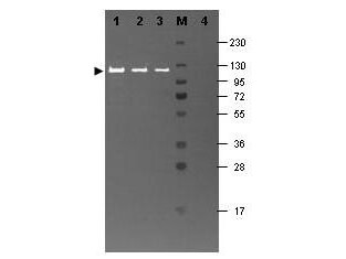
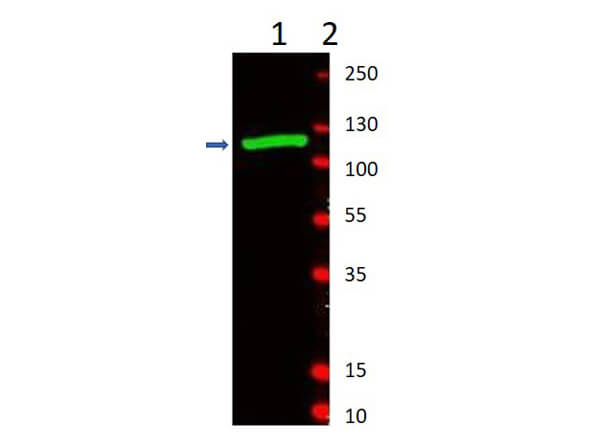
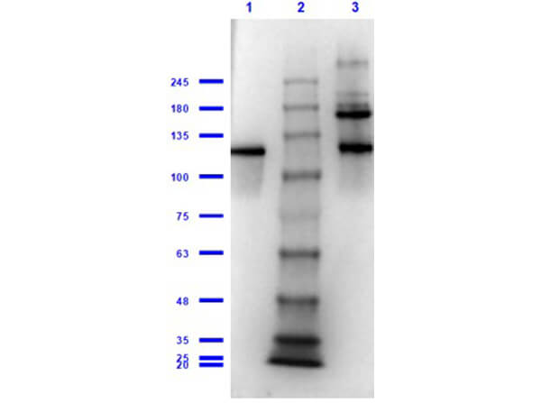
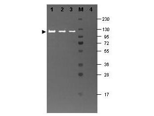
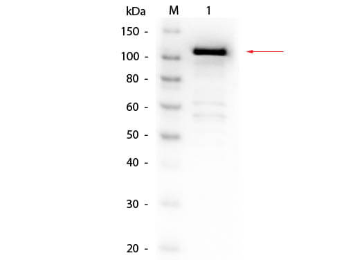
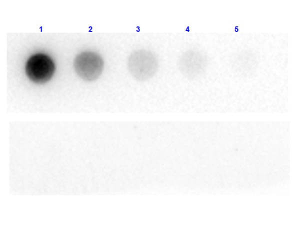
Anti-Beta-Gal Antibody has been tested by ELISA and western blot and is suitable for dot blot, immunofluorescence microscopy, immunoprecipitation, conjugation and most immunological methods requiring high titer and specificity. The antibody recognizes both frozen tissue sections, paraffin embedded tissue and 4% paraformaldehyde fixed tissue for most immunohistochemical analysis. A 1:1,500 dilution has been reported to detect beta-galactosidase in adult rat spinal cord tissue after infection with helper-dependent adenovirus expressing lacZ. In this particular experiment, tissue was perfused with 4% paraformaldehyde and cryostat-cut (35 µm) to produce free-floating sections.
Formulation
10.0 mg/mL by UV absorbance at 280 nm
0.02 M Potassium Phosphate, 0.15 M Sodium Chloride, pH 7.2
0.01% (w/v) Sodium Azide
None
1.0 mL
Restore with deionized water (or equivalent)
Shipping & Handling
Ambient
Store vial at 4° C prior to restoration. For extended storage aliquot contents and freeze at -20° C or below. Avoid cycles of freezing and thawing. Centrifuge product if not completely clear after standing at room temperature. This product is stable for several weeks at 4° C as an undiluted liquid. Dilute only prior to immediate use.
Expiration date is one (1) year from date of receipt.
Anti Beta Galactosidase Antibody recognizes the enzyme beta galactosidase, or β-galactosidase, that is a component of assays used frequently in genetics, molecular biology (see X-gal) for a blue white screen, and other life sciences. IPTG induces production of β-galactosidase by binding and inhibiting the lac repressor. Since it is highly expressed and accumulated in lysosomes in senescent cells, it is used as a senescence biomarker both in vivo and in vitro in qualitative and quantitative assays, despite its limitations. Anti-beta Galactosidase Antibody is ideal for investigators involved in enzyme research.
Krolewski RC et al. (2020). A Group of olfactory Receptor Alleles that encode full Length proteins are Down-Regulated as olfactory Sensory neurons Mature. Sci Rep.
Applications
IF, Confocal Microscopy
Pfannenstiel SC. et al. (2020). Hearing Preservation Following Repeated Adenovector Delivery. Anat Rec (Hoboken).
Applications
Undefined
Kodo K et al. (2017). Regulation of Sema3c and the interaction between cardiac neural crest and second heart field during outflow tract development. Sci Rep.
Applications
IHC, ICC, Histology; Multiplex Assay
El Agha E, Moiseenko A, Kheirollahi V, et al. (2017). Two-Way Conversion between Lipogenic and Myogenic Fibroblastic Phenotypes Marks the Progression and Resolution of Lung Fibrosis Cell Stem Cell.
Applications
IF, Confocal Microscopy; IHC, ICC, Histology
Yamanaka T et al. (2016). Differential roles of NF-Y transcription factor in ER chaperone expression and neuronal maintenance in the CNS. Sci Rep.
Applications
IF, Confocal Microscopy; Multiplex Assay
Ushijima T et al. (2014). CCAAT/enhancer-binding protein β regulates the repression of type II collagen expression during the differentiation from proliferative to hypertrophic chondrocytes. J Biol Chem
Applications
IHC, ICC, Histology
Yamanaka T et al. (2014). NF-Y inactivation causes atypical neurodegeneration characterized by ubiquitin and p62 accumulation and endoplasmic reticulum disorganization. Nat Commun.
Applications
IF, Confocal Microscopy; IHC, ICC, Histology; Multiplex Assay
Van Batavia J et al. (2014). Bladder cancers arise from distinct urothelial sub-populations. Nat Cell Biol.
Applications
IHC, ICC, Histology; Multiplex Assay
Yang X. et al. (2013). Simultaneous activation of Kras and inactivation of p53 induces soft tissue sarcoma and bladder urothelial hyperplasia. PLoS One
Applications
IF, Confocal Microscopy; Multiplex Assay
Yamanaka T et al. (2013). Loss of aPKCλ in differentiated neurons disrupts the polarity complex but does not induce obvious neuronal loss or disorientation in mouse brains. PLoS One
Applications
IHC, ICC, Histology
Hubener J et al. (2011). N-terminal ataxin-3 causes neurological symptoms with inclusions, endoplasmic reticulum stress and ribosomal dislocation. Brain.
Applications
IHC, ICC, Histology
Alaynick WA et al. (2009). ERRγ regulates cardiac, gastric, and renal potassium homeostasis. Mol Endocrinol.
Applications
IHC, ICC, Histology
Biju KC et al. (2008). Deletion of voltage-gated channel affects glomerular refinement and odorant receptor expression in the mouse olfactory system. J Comp Neurol.
Applications
IF, Confocal Microscopy; Multiplex Assay
Hayworth CR et al. (2006). Induction of neuregulin signaling in mouse schwann cells in vivo mimics responses to denervation. J Neurosci
Applications
IF, Confocal Microscopy
Nguyen HG et al. (2005). Conditional overexpression of transgenes in megakaryocytes and platelets in vivo. Blood
Applications
IF, Confocal Microscopy; WB, IB, PCA
Dufresne-Martin G et al. (2005). Peptide mass fingerprinting by matrix‐assisted laser desorption ionization mass spectrometry of proteins detected by immunostaining on nitrocellulose. Proteomics.
Applications
WB, IB, PCA
Young HE et al. (2004). Clonogenic analysis reveals reserve stem cells in postnatal mammals. II. Pluripotent epiblastic‐like stem cells. Anat Rec A Discov Mol Cell Evol Biol.
Applications
IF, Confocal Microscopy; Multiplex Assay
This product is for research use only and is not intended for therapeutic or diagnostic applications. Please contact a technical service representative for more information. All products of animal origin manufactured by Rockland Immunochemicals are derived from starting materials of North American origin. Collection was performed in United States Department of Agriculture (USDA) inspected facilities and all materials have been inspected and certified to be free of disease and suitable for exportation. All properties listed are typical characteristics and are not specifications. All suggestions and data are offered in good faith but without guarantee as conditions and methods of use of our products are beyond our control. All claims must be made within 30 days following the date of delivery. The prospective user must determine the suitability of our materials before adopting them on a commercial scale. Suggested uses of our products are not recommendations to use our products in violation of any patent or as a license under any patent of Rockland Immunochemicals, Inc. If you require a commercial license to use this material and do not have one, then return this material, unopened to: Rockland Inc., P.O. BOX 5199, Limerick, Pennsylvania, USA.

