AURORA KINASE B phospho T232 Antibody
Rabbit Polyclonal
35 References
600-401-677S
600-401-677
25 µL
100 µg
Liquid (sterile filtered)
Liquid (sterile filtered)
WB, ELISA, IHC, IF, Multiplex
Human, Monkey
Rabbit
Shipping info:
$50.00 to US & $70.00 to Canada for most products. Final costs are calculated at checkout.
Product Details
Anti-Aurora B pT232 (RABBIT) Antibody - 600-401-677
rabbit anti-Aurora B pT232 Antibody, Phospho Aurora B, AIK2 antibody, AIM1 antibody, ARK2 antibody, AurB antibody, AURKB antibody, Aurora 1 antibody, Aurora and Ipl1 like midbody associated protein 1 antibody
Rabbit
Polyclonal
IgG
Target Details
AURKB - View All AURKB Products
Human, Monkey
Phosphorylation
Conjugated Peptide
This affinity purified antibody was prepared from whole rabbit serum produced by repeated immunizations with a synthetic peptide corresponding to an internal region surrounding T232 of Human Aurora Kinase B protein.
Anti-Phospho Aurora B pT232 affinity purified antibody is directed against the phosphorylated form of human Aurora Kinase B at the pT232 residue. The product was affinity purified from monospecific antiserum by immunoaffinity purification. Antiserum was first purified against the phosphorylated form of the immunizing peptide. The resultant affinity purified antibody was then cross-adsorbed against the non-phosphorylated form of the immunizing peptide. Reactivity occurs against human Aurora Kinase B pT232 protein and the antibody is specific for the phosphorylated form of the protein. Reactivity with non-phosphorylated human Aurora Kinase B is minimal by ELISA. No reaction is expected against Aurora Kinase A. However, 100% sequence homology as indicated by BLAST analysis is on record for this protein from human, mouse, rat, cow, pig, dog and chimpanzee. Cross reactivity with Aurora Kinase B from other sources is not known.
Application Details
ELISA, IHC, WB
IF, Multiplex
- View References
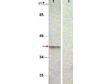
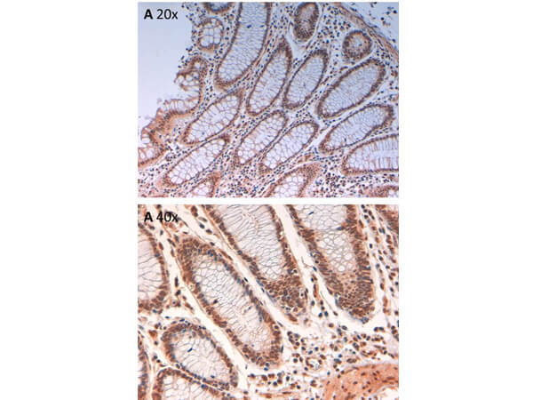
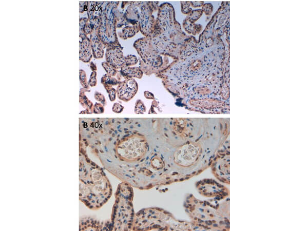
Phospho pT232 Aurora B antibody has been tested for use in ELISA, immunohistochemistry, and by western blot. See below for specific protocol. Expect a band approximately 39 kDa in size corresponding to Aurora Kinase B by western blotting in the appropriate cell lysate or extract. HeLa cell lysate can be used as a positive control.
Formulation
0.87 mg/mL by UV absorbance at 280 nm
0.02 M Potassium Phosphate, 0.15 M Sodium Chloride, pH 7.2
0.01% (w/v) Sodium Azide
None
Shipping & Handling
Dry Ice
Store Phospho Specific Antibody at -20° C prior to opening. Aliquot contents and freeze at -20° C or below for extended storage. Avoid cycles of freezing and thawing. Centrifuge product if not completely clear after standing at room temperature. This product is stable for several weeks at 4° C as an undiluted liquid. Dilute only prior to immediate use.
Expiration date is one (1) year from date of receipt.
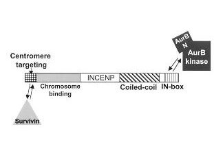
Aurora Kinase B (Aurora-B) is a Ser/Thr protein kinase member of the Aurora subfamily that may be directly involved in regulating the cleavage of polar spindle microtubules and is a key regulator for the onset of cytokinesis during mitosis. Aurora Kinase B is localized to the midzone of central spindle in late anaphase and concentrated into the midbody in telophase and cytokinesis and is colocalized with gamma tubulin in the mid-body. High levels of Aurora B expression are seen in the thymus, although it is also expressed in the spleen, lung, testis, colon, placenta and fetal liver. Aurora B is expressed during S and G2/M phase and expression is up-regulated in cancer cells during M phase. Anti-AUROA B pT232 Antibody is useful for researchers interested in gene expression, DNA damage, cytokinesis, and transcription activities.
Li J et al. (2024). METTL16-mediated N6-methyladenosine modification of Soga1 enables proper chromosome segregation and chromosomal stability in colorectal cancer. Cell Prolif.
Applications
IF, Confocal Microscopy
Roshan P et al. (2023). An Aurora B-RPA signaling axis secures chromosome segregation fidelity. Nat Commun.
Applications
WB, IB, PCA
Jiang H et al. (2023). Human Endonuclease ANKLE1 Localizes at the Midbody and Processes Chromatin Bridges to Prevent DNA Damage and cGAS-STING Activation. Adv Sci (Weinh).
Applications
IF, Confocal Microscopy
Kumar R et al. (2023). DENND2B activates Rab35 at the intercellular bridge, regulating cytokinetic abscission and tetraploidy. Cell Rep.
Applications
IF, Confocal Microscopy
Sobajima T et al. (2023). PP6 regulation of Aurora A-TPX2 limits NDC80 phosphorylation and mitotic spindle size. J Cell Biol.
Applications
IF, Confocal Microscopy; WB, IB, PCA
Song H et al. (2023). Hornerin mediates phosphorylation of the polo-box domain in Plk1 by Chk1 to induce death in mitosis. Cell Death Differ.
Applications
IF, Confocal Microscopy
Zhang M et al. (2022). SKAP interacts with Aurora B to guide end-on capture of spindle microtubules via phase separation. J Mol Cell Biol.
Applications
IF, Confocal Microscopy
LaJoie D et al. (2022). A role for Nup153 in nuclear assembly reveals differential requirements for targeting of nuclear envelope constituents. Mol Biol Cell.
Applications
IF, Confocal Microscopy
Zhang J et al. (2022). Topoisomerase II dysfunction causes metaphase I arrest by activating Aurora B, SAC and MPF and prevents PB1 abscission in mouse oocytes. Biol Reprod.
Applications
IF, Confocal Microscopy
Zhang X et al. (2021). Wip1 controls the translocation of the chromosomal passenger complex to the central spindle for faithful mitotic exit. Cell Mol Life Sci.
Applications
Undefined
Laine LJ et al. (2021). VTT-006, an anti-mitotic compound, binds to the Ndc80 complex and suppresses cancer cell growth in vitro. Oncoscience.
Applications
IF, Confocal Microscopy
Williams LK et al. (2021). Identification of abscission checkpoint bodies as structures that regulate ESCRT factors to control abscission timing. Elife
Applications
IF, Confocal Microscopy
Barroso-Vilares M et al. (2020). Small‐molecule inhibition of aging‐associated chromosomal instability delays cellular senescence. EMBO Rep.
Applications
IF, Confocal Microscopy
Sharp JA et al. (2020). Cell division requires RNA eviction from condensing chromosomes. J Cell Biol.
Applications
Undefined
Huang Y et al. (2019). BubR1 phosphorylates CENP-E as a switch enabling the transition from lateral association to end-on capture of spindle microtubules. Cell Res.
Applications
WB, IB, PCA
Achuthankutty D et al. (2019). Regulation of ETAA1-mediated ATR activation couples DNA replication fidelity and genome stability. J Cell Biol.
Applications
WB, IB, PCA
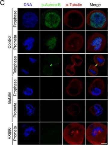
Xie et al. (2018). Cardiac glycoside bufalin blocks cancer cell growth by inhibition of Aurora A and Aurora B activation via PI3K-Akt pathway. Oncotarget
Applications
IF, Confocal Microscopy; WB, IB, PCA; Multiplex Assay
Skowyra et al. (2018). USP9X Limits Mitotic Checkpoint Complex Turnover to Strengthen the Spindle Assembly Checkpoint and Guard against Chromosomal Instability. Cell Reports
Applications
IF, Confocal Microscopy
Drpic et al. (2018). Chromosome Segregation Is Biased by Kinetochore Size. Current Biology
Applications
IF, Confocal Microscopy
Novais-Cruz et al. (2018). Mitotic progression, arrest, exit or death relies on centromere structural integrity, rather than de novo transcription. Elife
Applications
IF, Confocal Microscopy
de Carcer G et al. (2018). Plk1 overexpression induces chromosomal instability and suppresses tumor development. Nat Commun.
Applications
IF, Confocal Microscopy
Liu S et al. (2018). Nuclear envelope assembly defects link mitotic errors to chromothripsis. Nature.
Applications
IF, Confocal Microscopy
Weith M et al. (2018). Ubiquitin-independent disassembly by a p97 AAA-ATPase complex drives PP1 holoenzyme formation. Mol Cell.
Applications
IF, Confocal Microscopy
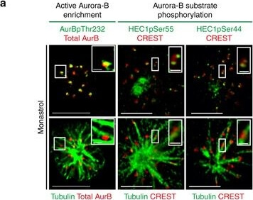
Shrestha et al. (2017). Aurora-B kinase pathway controls the lateral to end-on conversion of kinetochore-microtubule attachments in human cells. Nature Communications
Applications
IF, Confocal Microscopy; WB, IB, PCA; Multiplex Assay
Mo et al. (2016). Acetylation of Aurora B by TIP60 ensures accurate chromosomal segregation. Nature Chemical Biology
Applications
WB, IB, PCA
Redli et al. (2016). The Ska complex promotes Aurora B activity to ensure chromosome biorientation. Journal of Cell Biology
Applications
IF, Confocal Microscopy
Lee S et al. (2016). IK-guided PP2A suppresses Aurora B activity in the interphase of tumor cells. Cell Mol Life Sci.
Applications
IF, Confocal Microscopy; WB, IB, PCA
Saurin AT et al. (2016). Studying kinetochore kinases. Methods Mol Biol.
Applications
IF, Confocal Microscopy
Asghar et al. (2015). Bub1 autophosphorylation feeds back to regulate kinetochore docking and promote localized substrate phosphorylation. Nature Communications
Applications
IF, Confocal Microscopy
Nijenhuis W, Vallardi G, Teixeira A, Kops GJ, Saurin AT. (2014). Negative feedback at kinetochores underlies a responsive spindle checkpoint signal. Nat Cell Biol.
Applications
WB, IB, PCA; IF, Confocal Microscopy
Ohishi T et al. (2014). TRF1 ensures the centromeric function of Aurora-B and proper chromosome segregation. Mol Cell Biol.
Applications
IF, Confocal Microscopy
Thoresen SB et al. (2014). ANCHR mediates Aurora-B-dependent abscission checkpoint control through retention of VPS4. Nat Cell Biol.
Applications
IF, Confocal Microscopy
Carretero M et al. (2013). Pds5B is required for cohesion establishment and Aurora B accumulation at centromeres. EMBO J.
Applications
IF, Confocal Microscopy
Rivera T et al. (2012). Xenopus Shugoshin 2 regulates the spindle assembly pathway mediated by the chromosomal passenger complex. EMBO J.
Applications
IF, Confocal Microscopy
Mackay DR et al. (2010). Defects in nuclear pore assembly lead to activation of an Aurora B–mediated abscission checkpoint. J Cell Biol.
Applications
IF, Confocal Microscopy
This product is for research use only and is not intended for therapeutic or diagnostic applications. Please contact a technical service representative for more information. All products of animal origin manufactured by Rockland Immunochemicals are derived from starting materials of North American origin. Collection was performed in United States Department of Agriculture (USDA) inspected facilities and all materials have been inspected and certified to be free of disease and suitable for exportation. All properties listed are typical characteristics and are not specifications. All suggestions and data are offered in good faith but without guarantee as conditions and methods of use of our products are beyond our control. All claims must be made within 30 days following the date of delivery. The prospective user must determine the suitability of our materials before adopting them on a commercial scale. Suggested uses of our products are not recommendations to use our products in violation of any patent or as a license under any patent of Rockland Immunochemicals, Inc. If you require a commercial license to use this material and do not have one, then return this material, unopened to: Rockland Inc., P.O. BOX 5199, Limerick, Pennsylvania, USA.

