GFP Chemiluminescent Western Blot Kit
KCA215
1 Kit
n/a
WB
GFP, eGFP, rGFP
Shipping info:
$50.00 to US & $70.00 to Canada for most products. Final costs are calculated at checkout.
Product Details
GFP Western Blot Kit: for GFP Chemiluminescent Western Blotting - KCA215
GFP Western Blotting Kit, GFP Chemiluminescent Kit, Green Fluorescent Protein Antibody Kit, Immunoblotting Kit, GFP, rGFP, eGFP
Monoclonal
Chemiluminescent Western Blot Kit
Target Details
GFP, eGFP, rGFP
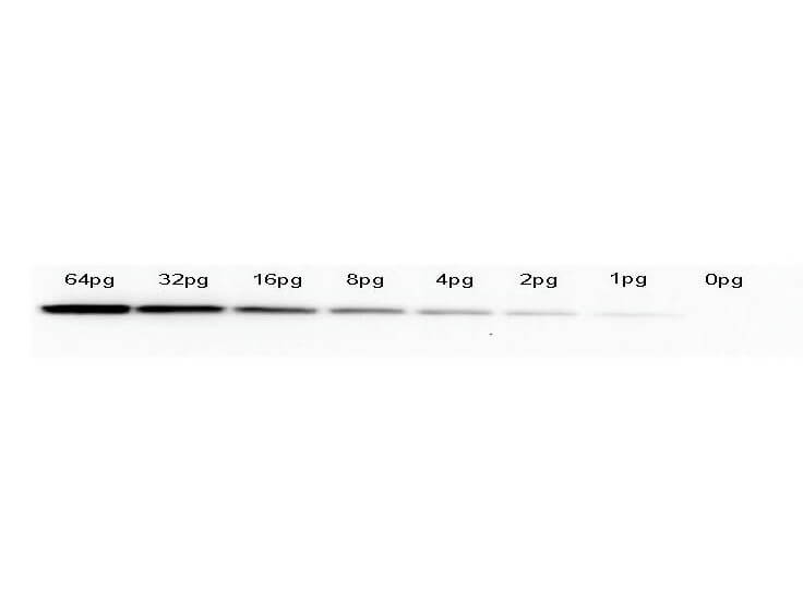
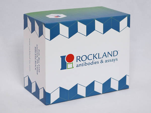
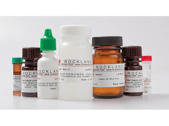
Rockland Immunochemicals’ Chemiluminescent Western Blot Kit for GFP combines all of the necessary reagents with a rapid proven protocol and the extremely high signal detection of our luminol chemiluminescent substrate for the detection of recombinant proteins containing GFP and its variants. The Chemiluminescent Western Blot Kit design includes straightforward procedures and color-coding to allow for ease of use. This kit contains: GFP protein, Mouse IgG control, mouse anti-GFP antibody, anti-mouse IgG peroxidase, Tween-20, BSA, and Femtomax, along with additional supplies and protocol; sufficient substrate for up to 30 mini blots at 7.5 x 8 cm2 (1,800 cm2) and is stable for at least 1 year when stored as indicated.
Application Details
WB
GFP Western Blot Kit is ideal for detection of GFP-tagged recombinant proteins by western blot. The GFP Western Blot kit makes use of Rockland's optimized anti-GFP antibody. The Chemiluminescent GFP Western Blot kit is useful for both western blotting and dot blotting methods. Please read the entire product insert prior to use.
Formulation
Wash buffers MUST NOT contain SODIUM AZIDE or other inhibitors of peroxidase activity!
Shipping & Handling
Wet Ice
See GFP Western Blot kit insert for complete instructions.
See kit insert for complete instructions.
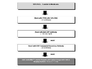
GFP Chemiluminescent Western Blot Kit allows for the detection of GFP-tagged recombinant proteins present in cell lysates provided by the user. After protein separation by SDS-PAGE and transfer, the membrane is probed with monoclonal Anti-GFP. Detection of the membrane bound antibody-antigen complex is achieved by the addition of a secondary antibody conjugated to the enzyme horseradish peroxidase. The enzyme reacts with a specialized formulation of luminol, an extremely sensitive, non-radioactive substrate that emits light and allows visualization using X-ray film or other imaging methods, including highly sensitive CCD cameras and imaging systems.
This product is for research use only and is not intended for therapeutic or diagnostic applications. Please contact a technical service representative for more information. All products of animal origin manufactured by Rockland Immunochemicals are derived from starting materials of North American origin. Collection was performed in United States Department of Agriculture (USDA) inspected facilities and all materials have been inspected and certified to be free of disease and suitable for exportation. All properties listed are typical characteristics and are not specifications. All suggestions and data are offered in good faith but without guarantee as conditions and methods of use of our products are beyond our control. All claims must be made within 30 days following the date of delivery. The prospective user must determine the suitability of our materials before adopting them on a commercial scale. Suggested uses of our products are not recommendations to use our products in violation of any patent or as a license under any patent of Rockland Immunochemicals, Inc. If you require a commercial license to use this material and do not have one, then return this material, unopened to: Rockland Inc., P.O. BOX 5199, Limerick, Pennsylvania, USA.


