Immunoassay Development
Immunoassay Development
Advance confidently with immunoassay development and implementation services from the antibody experts at Rockland. Through decades of experience developing and running thousands of immunoassays to ensure the quality of our products and support our service customers, we have optimized processes that maximize success.
Our custom immunoassay services include development, qualification, validation, and implementation of Western blot- and ELISA-based assays—including in-cell Westerns and cell-based ELISAs—that support a range of applications, from discovery research and pre-clinical studies to bioprocess monitoring and lot release testing and more.
Immunoassay Capabilities
Whether you need a Western blot or an ELISA, we can develop and run an immunoassay using many of the most widely used chemiluminescence and fluorescence detection technologies, including:
Example Immunoassays
Here are just a few examples of the types of immunoassays we can develop and run.

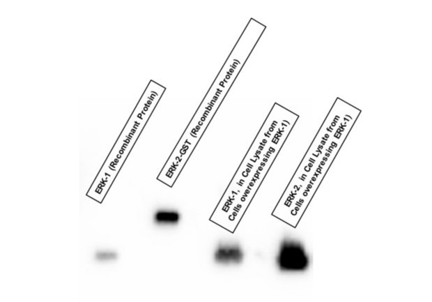
Assessing antibody specificity and cross-reactivity by Western blot
Here we developed a Western blot assay to assess the specificity and cross-reactivity of an antibody raised against ERK-2 to ERK-1 proteins.

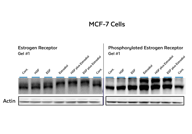
Identifying molecular changes in neoplastic cells by Western blot
In this Western blot electrophoretic mobility shift assay (EMSA), we developed a way to assess estradiol-stimulated phosphorylation of estrogen receptors on MCF-7 breast cancer cells. Phosphorylation is detected as the upward shift of the estrogen receptor band when probed with an antibody directed against the estrogen receptor. The Western blot membrane on the right was probed with an antibody directed against the phosphorylated estrogen receptor.

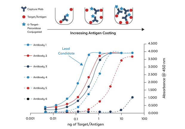
Comparison of Capture Antibodies by Direct ELISA
Plates were coated with a serial dilution of antigen and probed with several capture antibodies. The lead capture antibody has a limit of detection below 100 pg.

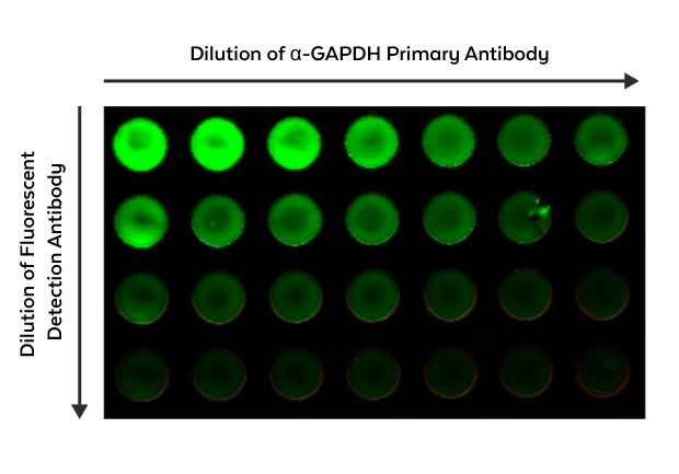
Cell-based Fluorescent ELISA
Adherent epithelial cells were fixed onto a plate and probed with primary antibody anti-GAPDH and detected by a secondary DyLight 488 coupled antibody.
Interested in immunoassay development & implementation?
We can help move your program forward—contact us to find out how.